Yiyuan Zhu , Enbo Xu , Jun Yin , Weidong Xu , Donghong Liu
{"title":"Visualization, modeling and analysis of salmon muscle structure: Based on micro-CT","authors":"Yiyuan Zhu , Enbo Xu , Jun Yin , Weidong Xu , Donghong Liu","doi":"10.1016/j.foostr.2023.100325","DOIUrl":null,"url":null,"abstract":"<div><p><span>Seafood analogs using culture cells or plant-based materials are rising for future food supply, but their </span>bionic design is still an issue on finely simulating the multiscale structure and function of fishes. For salmon, we compared the back, belly and tail tissues via micro computed tomography (micro-CT) scanning with optimized tissue contrast and spatial resolution. The optimum staining conditions of salmon muscle tissue for micro-CT was 3.75 % IKI (iodine-potassium iodide) solution for 7 days, and the muscle could be separated well from fat and connective tissue. The results of visualization analysis were correlated with the texture profile analysis. A relatively high magnification (3 µm/pixel) was used with fiber tracing procedure to analyze the connection of muscle fiber bundles surrounded by soft tissue. Single muscle fiber (e.g., diameter 117 µm) was successfully and artificially separated from whole visualized tissue through manual division. Overall, this study showed a novel ultra-high-resolution method based on micro-CT to conduct digital analysis of muscle fascicle architecture, and constructed printable model of bionic animal/plant tissue for food design.</p></div>","PeriodicalId":48640,"journal":{"name":"Food Structure-Netherlands","volume":"37 ","pages":"Article 100325"},"PeriodicalIF":5.9000,"publicationDate":"2023-07-01","publicationTypes":"Journal Article","fieldsOfStudy":null,"isOpenAccess":false,"openAccessPdf":"","citationCount":"0","resultStr":null,"platform":"Semanticscholar","paperid":null,"PeriodicalName":"Food Structure-Netherlands","FirstCategoryId":"97","ListUrlMain":"https://www.sciencedirect.com/science/article/pii/S2213329123000187","RegionNum":3,"RegionCategory":"农林科学","ArticlePicture":[],"TitleCN":null,"AbstractTextCN":null,"PMCID":null,"EPubDate":"","PubModel":"","JCR":"Q1","JCRName":"FOOD SCIENCE & TECHNOLOGY","Score":null,"Total":0}
引用次数: 0
Abstract
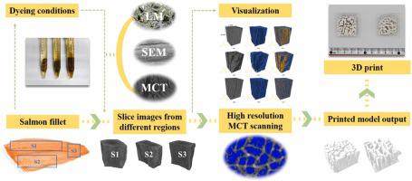
Seafood analogs using culture cells or plant-based materials are rising for future food supply, but their bionic design is still an issue on finely simulating the multiscale structure and function of fishes. For salmon, we compared the back, belly and tail tissues via micro computed tomography (micro-CT) scanning with optimized tissue contrast and spatial resolution. The optimum staining conditions of salmon muscle tissue for micro-CT was 3.75 % IKI (iodine-potassium iodide) solution for 7 days, and the muscle could be separated well from fat and connective tissue. The results of visualization analysis were correlated with the texture profile analysis. A relatively high magnification (3 µm/pixel) was used with fiber tracing procedure to analyze the connection of muscle fiber bundles surrounded by soft tissue. Single muscle fiber (e.g., diameter 117 µm) was successfully and artificially separated from whole visualized tissue through manual division. Overall, this study showed a novel ultra-high-resolution method based on micro-CT to conduct digital analysis of muscle fascicle architecture, and constructed printable model of bionic animal/plant tissue for food design.
期刊介绍:
Food Structure is the premier international forum devoted to the publication of high-quality original research on food structure. The focus of this journal is on food structure in the context of its relationship with molecular composition, processing and macroscopic properties (e.g., shelf stability, sensory properties, etc.). Manuscripts that only report qualitative findings and micrographs and that lack sound hypothesis-driven, quantitative structure-function research are not accepted. Significance of the research findings for the food science community and/or industry must also be highlighted.


 分享
分享
 求助内容:
求助内容: 应助结果提醒方式:
 扫码关注我们
