No evidence of increased cerebrovascular involvement in adult neurologically-asymptomatic β-Thalassaemia. A multicentre multimodal magnetic resonance study
Immacolata Tartaglione, Camilla Russo, Andrea Elefante, Martina Caiazza, Maddalena Casale, Rosanna Di Concilio, Angela Ciancio, Elisa De Michele, Giovanni Amendola, Paolo Gritti, Pasquale A. Carafa, Teresa Ferrantino, Antonella Centanni, Noemi Ippolito, Violetta Caserta, Tiziana Oliveto, Ilaria Granato, Gianluca Femina, Fabrizio Esposito, Sara Ponticorvo, Andrea G. Russo, Antonietta Canna, Mario Ermani, Mario Cirillo, Silverio Perrotta, Renzo Manara
{"title":"No evidence of increased cerebrovascular involvement in adult neurologically-asymptomatic β-Thalassaemia. A multicentre multimodal magnetic resonance study","authors":"Immacolata Tartaglione, Camilla Russo, Andrea Elefante, Martina Caiazza, Maddalena Casale, Rosanna Di Concilio, Angela Ciancio, Elisa De Michele, Giovanni Amendola, Paolo Gritti, Pasquale A. Carafa, Teresa Ferrantino, Antonella Centanni, Noemi Ippolito, Violetta Caserta, Tiziana Oliveto, Ilaria Granato, Gianluca Femina, Fabrizio Esposito, Sara Ponticorvo, Andrea G. Russo, Antonietta Canna, Mario Ermani, Mario Cirillo, Silverio Perrotta, Renzo Manara","doi":"10.1111/bjh.15834","DOIUrl":null,"url":null,"abstract":"<p>Multi-factorial causes jeopardize brain integrity in β-thalassaemia. Intracranial parenchymal and vascular changes have been reported among young β-thalassaemia patients but conventional magnetic resonance imaging (MRI) findings are contradictory making early MRI and magnetic resonance angiography (MRA)/venography monitoring a matter of debate.</p><p>This study prospectively investigated 75 neurologically asymptomatic β-thalassaemia patients (mean-age 35·2 ± 10·7 years; 52/75 transfusion-dependent; 41/75 splenectomised) using a 3T magnetic resonance scanner; clinical, laboratory and treatment data were also collected. White matter ischaemic-like abnormalities, intracranial artery stenoses, aneurysms and sinus venous thrombosis were compared between patients and 56 healthy controls (mean-age 33·9 ± 10·8 years). No patient or control showed silent territorial or lacunar strokes, intracranial artery stenoses or signs of sinus thrombosis. White matter lesions were found both in patients (35/75, 46·7%) and controls (28/56, 50·0%), without differences in terms of number (4·0 ± 10·6 vs. 4·6 ± 9·1, <i>P</i> = 0·63), size and Fazekas’ Score. Intracranial aneurysms did not differ between patients and controls for incidence rate (7/75, 9·3% vs. 5/56, 8·9%), size and site. Vascular and parenchymal abnormality rate did not differ according to treatments or clinical phenotype. According to this study, asymptomatic β-thalassaemia patients treated according to current guidelines do not seem to carry an increased risk of brain and intracranial vascular changes, thus weakening recommendations for regular brain MRI monitoring.</p>","PeriodicalId":135,"journal":{"name":"British Journal of Haematology","volume":"185 4","pages":"733-742"},"PeriodicalIF":3.8000,"publicationDate":"2019-03-05","publicationTypes":"Journal Article","fieldsOfStudy":null,"isOpenAccess":false,"openAccessPdf":"https://sci-hub-pdf.com/10.1111/bjh.15834","citationCount":"13","resultStr":null,"platform":"Semanticscholar","paperid":null,"PeriodicalName":"British Journal of Haematology","FirstCategoryId":"3","ListUrlMain":"https://onlinelibrary.wiley.com/doi/10.1111/bjh.15834","RegionNum":2,"RegionCategory":"医学","ArticlePicture":[],"TitleCN":null,"AbstractTextCN":null,"PMCID":null,"EPubDate":"","PubModel":"","JCR":"Q1","JCRName":"HEMATOLOGY","Score":null,"Total":0}
引用次数: 13
Abstract
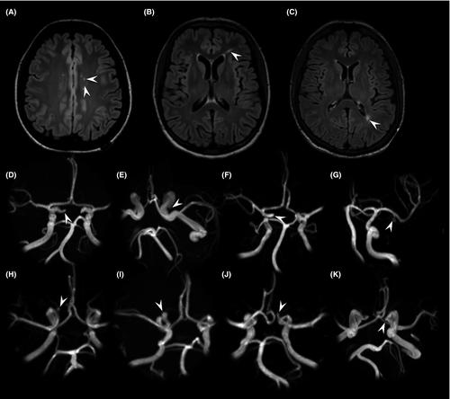
Multi-factorial causes jeopardize brain integrity in β-thalassaemia. Intracranial parenchymal and vascular changes have been reported among young β-thalassaemia patients but conventional magnetic resonance imaging (MRI) findings are contradictory making early MRI and magnetic resonance angiography (MRA)/venography monitoring a matter of debate.
This study prospectively investigated 75 neurologically asymptomatic β-thalassaemia patients (mean-age 35·2 ± 10·7 years; 52/75 transfusion-dependent; 41/75 splenectomised) using a 3T magnetic resonance scanner; clinical, laboratory and treatment data were also collected. White matter ischaemic-like abnormalities, intracranial artery stenoses, aneurysms and sinus venous thrombosis were compared between patients and 56 healthy controls (mean-age 33·9 ± 10·8 years). No patient or control showed silent territorial or lacunar strokes, intracranial artery stenoses or signs of sinus thrombosis. White matter lesions were found both in patients (35/75, 46·7%) and controls (28/56, 50·0%), without differences in terms of number (4·0 ± 10·6 vs. 4·6 ± 9·1, P = 0·63), size and Fazekas’ Score. Intracranial aneurysms did not differ between patients and controls for incidence rate (7/75, 9·3% vs. 5/56, 8·9%), size and site. Vascular and parenchymal abnormality rate did not differ according to treatments or clinical phenotype. According to this study, asymptomatic β-thalassaemia patients treated according to current guidelines do not seem to carry an increased risk of brain and intracranial vascular changes, thus weakening recommendations for regular brain MRI monitoring.
期刊介绍:
The British Journal of Haematology publishes original research papers in clinical, laboratory and experimental haematology. The Journal also features annotations, reviews, short reports, images in haematology and Letters to the Editor.


 分享
分享
 求助内容:
求助内容: 应助结果提醒方式:
 扫码关注我们
