Giovanni Paolino, Matteo Riccardo Di Nicola, Marina Yarygina, Carlo Mattozzi, Eduardo Quaranta, Vittoria Giulia Bianchi, Michele Donati, Santo Raffaele Mercuri
{"title":"Exclusive and Solitary Facial Porokeratosis: Pathogenesis and Literature Reappraisal of a Rare Entity.","authors":"Giovanni Paolino, Matteo Riccardo Di Nicola, Marina Yarygina, Carlo Mattozzi, Eduardo Quaranta, Vittoria Giulia Bianchi, Michele Donati, Santo Raffaele Mercuri","doi":"10.1159/000530936","DOIUrl":null,"url":null,"abstract":"<p><p>Porokeratosis is a group of well-known clinically distinct entities, characterised by different clinical aspects, but sharing a single common histological aspect, namely the cornoid lamella. Usually, porokeratosis occurs in the limbs and trunk, while it rarely involves the face, especially as an exclusive, single, and solitary lesion. We report the case of a 52-year-old Caucasian woman, with an 11-month history of a 2-cm slowly growing solitary, keratotic lesion on her left cheekbone. The patient did not present other cutaneous lesions on the face, as well as in other body sites. A cutaneous biopsy showed epidermal hyperplasia with multiple, sharply defined cornoid lamella, associated with an underlying attenuation of the granular layer and scattered dyskeratotic cells in the spinous layer. The superficial dermis underneath showed a mild lymphocytic infiltrate and fibrosis with remodelled collagen bundles. A final diagnosis of solitary facial porokeratosis was made.</p>","PeriodicalId":9619,"journal":{"name":"Case Reports in Dermatology","volume":"15 1","pages":"147-151"},"PeriodicalIF":0.8000,"publicationDate":"2023-09-05","publicationTypes":"Journal Article","fieldsOfStudy":null,"isOpenAccess":false,"openAccessPdf":"https://www.ncbi.nlm.nih.gov/pmc/articles/PMC10601618/pdf/","citationCount":"0","resultStr":null,"platform":"Semanticscholar","paperid":null,"PeriodicalName":"Case Reports in Dermatology","FirstCategoryId":"1085","ListUrlMain":"https://doi.org/10.1159/000530936","RegionNum":0,"RegionCategory":null,"ArticlePicture":[],"TitleCN":null,"AbstractTextCN":null,"PMCID":null,"EPubDate":"2023/1/1 0:00:00","PubModel":"eCollection","JCR":"Q4","JCRName":"DERMATOLOGY","Score":null,"Total":0}
引用次数: 0
Abstract
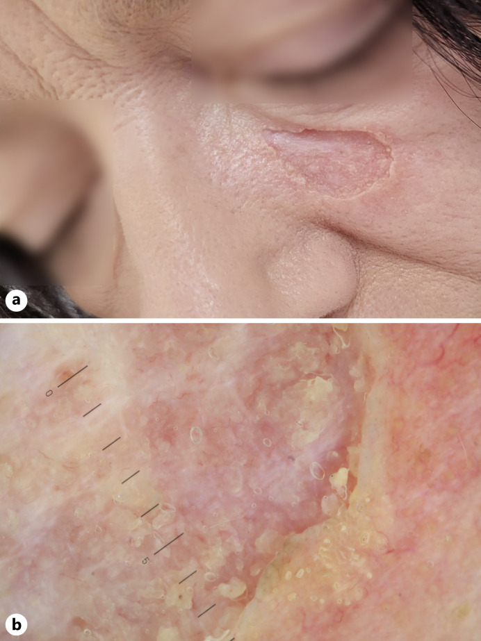
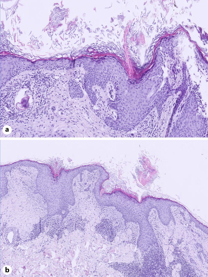
Porokeratosis is a group of well-known clinically distinct entities, characterised by different clinical aspects, but sharing a single common histological aspect, namely the cornoid lamella. Usually, porokeratosis occurs in the limbs and trunk, while it rarely involves the face, especially as an exclusive, single, and solitary lesion. We report the case of a 52-year-old Caucasian woman, with an 11-month history of a 2-cm slowly growing solitary, keratotic lesion on her left cheekbone. The patient did not present other cutaneous lesions on the face, as well as in other body sites. A cutaneous biopsy showed epidermal hyperplasia with multiple, sharply defined cornoid lamella, associated with an underlying attenuation of the granular layer and scattered dyskeratotic cells in the spinous layer. The superficial dermis underneath showed a mild lymphocytic infiltrate and fibrosis with remodelled collagen bundles. A final diagnosis of solitary facial porokeratosis was made.


 分享
分享
 求助内容:
求助内容: 应助结果提醒方式:
 扫码关注我们
