Jun Ho Choi, Soo Hyuk Lee, Jae Ha Hwang, Kwang Seog Kim, Sam Yong Lee
{"title":"Solitary fibrous tumor in the temporalis muscle: a case report and literature review.","authors":"Jun Ho Choi, Soo Hyuk Lee, Jae Ha Hwang, Kwang Seog Kim, Sam Yong Lee","doi":"10.7181/acfs.2023.00199","DOIUrl":null,"url":null,"abstract":"<p><p>Solitary fibrous tumor (SFT) is an infrequently occurring neoplasm most commonly observed in the pleura, but it can develop in the head and neck region in occasional cases. However, no reports have described SFT in the temporalis muscle. Herein, we present the first known case of SFT in the temporalis muscle. A 47-year-old man complained of a painless palpable mass on his right temple. Facial enhanced computed tomography identified a 4.0× 2.9× 1.4 cm mass presenting as a vascular tumor in the right temporalis muscle under the zygomatic arch. The mass was excised from the right temporalis muscle under general anesthesia. A histopathologic examination revealed that the mass was an SFT. No complications occurred after surgery, including functional disability or sensory loss. The patient was followed up for 3 months without complications. Although SFT in extrapulmonary regions is rare, it should be considered in the differential diagnosis of masses that occur in the temporal area.</p>","PeriodicalId":52238,"journal":{"name":"Archives of Craniofacial Surgery","volume":"24 5","pages":"230-235"},"PeriodicalIF":0.0000,"publicationDate":"2023-10-01","publicationTypes":"Journal Article","fieldsOfStudy":null,"isOpenAccess":false,"openAccessPdf":"https://www.ncbi.nlm.nih.gov/pmc/articles/PMC10622951/pdf/","citationCount":"0","resultStr":null,"platform":"Semanticscholar","paperid":null,"PeriodicalName":"Archives of Craniofacial Surgery","FirstCategoryId":"1085","ListUrlMain":"https://doi.org/10.7181/acfs.2023.00199","RegionNum":0,"RegionCategory":null,"ArticlePicture":[],"TitleCN":null,"AbstractTextCN":null,"PMCID":null,"EPubDate":"2023/10/20 0:00:00","PubModel":"Epub","JCR":"Q2","JCRName":"Medicine","Score":null,"Total":0}
引用次数: 0
Abstract
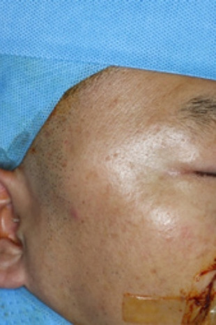
Solitary fibrous tumor (SFT) is an infrequently occurring neoplasm most commonly observed in the pleura, but it can develop in the head and neck region in occasional cases. However, no reports have described SFT in the temporalis muscle. Herein, we present the first known case of SFT in the temporalis muscle. A 47-year-old man complained of a painless palpable mass on his right temple. Facial enhanced computed tomography identified a 4.0× 2.9× 1.4 cm mass presenting as a vascular tumor in the right temporalis muscle under the zygomatic arch. The mass was excised from the right temporalis muscle under general anesthesia. A histopathologic examination revealed that the mass was an SFT. No complications occurred after surgery, including functional disability or sensory loss. The patient was followed up for 3 months without complications. Although SFT in extrapulmonary regions is rare, it should be considered in the differential diagnosis of masses that occur in the temporal area.


 分享
分享
 求助内容:
求助内容: 应助结果提醒方式:
 扫码关注我们
