Almoatazbellah Youssef, Andreas Rosenwald, Mathias Tillmann Rosenfeldt
{"title":"TelePi: an affordable telepathology microscope camera system anyone can build and use.","authors":"Almoatazbellah Youssef, Andreas Rosenwald, Mathias Tillmann Rosenfeldt","doi":"10.1007/s00428-023-03685-5","DOIUrl":null,"url":null,"abstract":"<p><p>Telepathology facilitates histological diagnoses through sharing expertise between pathologists. However, the associated costs are high and frequently prohibitive, especially in low-resource settings, where telepathology would paradoxically be of paramount importance due to a paucity of pathologists.We have constructed a telepathology system (TelePi) with a budget of < €120 using the small, single-board computer Raspberry Pi Zero and its High-Quality Camera Module in conjunction with a standard microscope and open-source software. The system requires no maintenance costs or service contracts, has a small footprint, can be moved and shared across several microscopes, and is independent from other computer operating systems. TelePi uses a responsive and high-resolution web-based live stream which allows remote consultation between two or more locations. TelePi can serve as a telepathology system for remote diagnostics of frozen sections. Additionally, it can be used as a standard microscope camera for teaching of medical students and for basic research. The quality of the TelePi system compared favorable to a commercially available telepathology system that exceed its cost by more than 125-fold. Additionally, still images are of publication quality equal to that of a whole slide scanner that costs 800 times more.In summary, TelePi is an affordable, versatile, and inexpensive camera system that potentially enables telepathology in low-resource settings without sacrificing image quality.</p>","PeriodicalId":23514,"journal":{"name":"Virchows Archiv","volume":" ","pages":"115-122"},"PeriodicalIF":3.1000,"publicationDate":"2024-07-01","publicationTypes":"Journal Article","fieldsOfStudy":null,"isOpenAccess":false,"openAccessPdf":"https://www.ncbi.nlm.nih.gov/pmc/articles/PMC11271423/pdf/","citationCount":"0","resultStr":null,"platform":"Semanticscholar","paperid":null,"PeriodicalName":"Virchows Archiv","FirstCategoryId":"3","ListUrlMain":"https://doi.org/10.1007/s00428-023-03685-5","RegionNum":3,"RegionCategory":"医学","ArticlePicture":[],"TitleCN":null,"AbstractTextCN":null,"PMCID":null,"EPubDate":"2023/11/7 0:00:00","PubModel":"Epub","JCR":"Q1","JCRName":"PATHOLOGY","Score":null,"Total":0}
引用次数: 0
Abstract
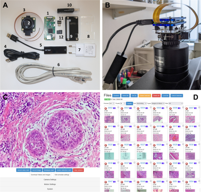
Telepathology facilitates histological diagnoses through sharing expertise between pathologists. However, the associated costs are high and frequently prohibitive, especially in low-resource settings, where telepathology would paradoxically be of paramount importance due to a paucity of pathologists.We have constructed a telepathology system (TelePi) with a budget of < €120 using the small, single-board computer Raspberry Pi Zero and its High-Quality Camera Module in conjunction with a standard microscope and open-source software. The system requires no maintenance costs or service contracts, has a small footprint, can be moved and shared across several microscopes, and is independent from other computer operating systems. TelePi uses a responsive and high-resolution web-based live stream which allows remote consultation between two or more locations. TelePi can serve as a telepathology system for remote diagnostics of frozen sections. Additionally, it can be used as a standard microscope camera for teaching of medical students and for basic research. The quality of the TelePi system compared favorable to a commercially available telepathology system that exceed its cost by more than 125-fold. Additionally, still images are of publication quality equal to that of a whole slide scanner that costs 800 times more.In summary, TelePi is an affordable, versatile, and inexpensive camera system that potentially enables telepathology in low-resource settings without sacrificing image quality.
期刊介绍:
Manuscripts of original studies reinforcing the evidence base of modern diagnostic pathology, using immunocytochemical, molecular and ultrastructural techniques, will be welcomed. In addition, papers on critical evaluation of diagnostic criteria but also broadsheets and guidelines with a solid evidence base will be considered. Consideration will also be given to reports of work in other fields relevant to the understanding of human pathology as well as manuscripts on the application of new methods and techniques in pathology. Submission of purely experimental articles is discouraged but manuscripts on experimental work applicable to diagnostic pathology are welcomed. Biomarker studies are welcomed but need to abide by strict rules (e.g. REMARK) of adequate sample size and relevant marker choice. Single marker studies on limited patient series without validated application will as a rule not be considered. Case reports will only be considered when they provide substantial new information with an impact on understanding disease or diagnostic practice.


 分享
分享
 求助内容:
求助内容: 应助结果提醒方式:
 扫码关注我们
