Narges Hassanpoor, Vahid Abdolrahimi, Mohamad Reza Niyousha
{"title":"Central Retinal Vein Occlusion with Three-Retinal Quadrant Involvement: Another Focus on Optic Disc Head Vascular Anatomy Variations.","authors":"Narges Hassanpoor, Vahid Abdolrahimi, Mohamad Reza Niyousha","doi":"10.1155/2023/6648367","DOIUrl":null,"url":null,"abstract":"<p><p>A 50-year-old male patient with sudden visual acuity loss in his right eye came to our clinic. Visual acuity at presentation was 1/10 in right eye and 10/10 in left. The patient was otherwise healthy Caucasian man without any history of previous systemic or ophthalmic disease. There was not any history of amblyopia and refractive error. Anterior segment findings were unremarkable. Three quadrants of retina were fully involved with central retinal vein occlusion (CRVO) features including retinal hemorrhages, retinal edema obscuring retinal details, and cotton wool spots while sparing inferior temporal quadrant. Inferior temporal quadrant sparing in this patient is due to a specific retinal vascular anatomical variation. In conclusion, in unusual presentations of retinal vascular branch obstructions, considering retinal vascular anatomy variations would help us to explain the clinical presentation more precisely in some cases.</p>","PeriodicalId":9603,"journal":{"name":"Case Reports in Ophthalmological Medicine","volume":"2023 ","pages":"6648367"},"PeriodicalIF":0.4000,"publicationDate":"2023-10-31","publicationTypes":"Journal Article","fieldsOfStudy":null,"isOpenAccess":false,"openAccessPdf":"https://www.ncbi.nlm.nih.gov/pmc/articles/PMC10630022/pdf/","citationCount":"0","resultStr":null,"platform":"Semanticscholar","paperid":null,"PeriodicalName":"Case Reports in Ophthalmological Medicine","FirstCategoryId":"1085","ListUrlMain":"https://doi.org/10.1155/2023/6648367","RegionNum":0,"RegionCategory":null,"ArticlePicture":[],"TitleCN":null,"AbstractTextCN":null,"PMCID":null,"EPubDate":"2023/1/1 0:00:00","PubModel":"eCollection","JCR":"Q4","JCRName":"OPHTHALMOLOGY","Score":null,"Total":0}
引用次数: 0
Abstract
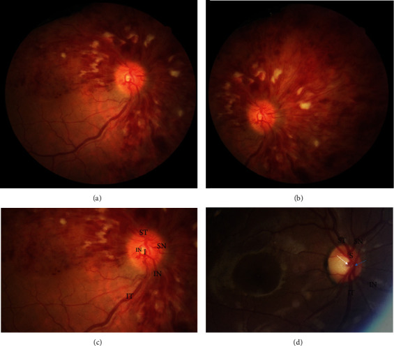
A 50-year-old male patient with sudden visual acuity loss in his right eye came to our clinic. Visual acuity at presentation was 1/10 in right eye and 10/10 in left. The patient was otherwise healthy Caucasian man without any history of previous systemic or ophthalmic disease. There was not any history of amblyopia and refractive error. Anterior segment findings were unremarkable. Three quadrants of retina were fully involved with central retinal vein occlusion (CRVO) features including retinal hemorrhages, retinal edema obscuring retinal details, and cotton wool spots while sparing inferior temporal quadrant. Inferior temporal quadrant sparing in this patient is due to a specific retinal vascular anatomical variation. In conclusion, in unusual presentations of retinal vascular branch obstructions, considering retinal vascular anatomy variations would help us to explain the clinical presentation more precisely in some cases.


 分享
分享
 求助内容:
求助内容: 应助结果提醒方式:
 扫码关注我们
