Ane Escobar, Mariana R Carvalho, Tiago H Silva, Rui L Reis, J Miguel Oliveira
{"title":"Longitudinally aligned inner-patterned silk fibroin conduits for peripheral nerve regeneration.","authors":"Ane Escobar, Mariana R Carvalho, Tiago H Silva, Rui L Reis, J Miguel Oliveira","doi":"10.1007/s44164-023-00050-3","DOIUrl":null,"url":null,"abstract":"<p><p>Peripheral nerve injuries represent a major clinical challenge, if nerve ends retract, there is no spontaneous regeneration, and grafts are required to proximate the nerve ends and give continuity to the nerve. The nerve guidance conduits (NGCs) presented in this work are silk fibroin (SF)-based, which is biocompatible and very versatile. The formation of conduits is obtained by forming a covalently cross-linked hydrogel in two concentric moulds, and the inner longitudinally aligned pattern of the SF NGCs is obtained through the use of a patterned inner mould. SF NGCs with two wall thicknesses of ~ 200 to ~ 400 μm are synthesized. Their physicochemical and mechanical characteristics have shown improved properties when the wall thickness is thicker such as resistance to kinking, which is of special importance as conduits might also be used to substitute nerves in flexible body parts. The Young modulus is higher for conduits with inner pattern, and none of the conduits has shown any salt deposition in presence of simulated body fluid, meaning they do not calcify; thus, the regeneration does not get impaired when conduits have contact with body fluids. In vitro studies demonstrated the biocompatibility of the SF NGCs; proliferation is enhanced when iSCs are cultured on top of conduits with longitudinally aligned pattern. BJ fibroblasts cannot infiltrate through the SF wall, avoiding scar tissue formation on the lumen of the graft when used in vivo. These conduits have been demonstrated to be very versatile and fulfil with the requirements for their use in PNR.</p><p><strong>Supplementary information: </strong>The online version contains supplementary material available at 10.1007/s44164-023-00050-3.</p>","PeriodicalId":73357,"journal":{"name":"In vitro models","volume":"18 1","pages":"195-205"},"PeriodicalIF":2.4000,"publicationDate":"2023-04-18","publicationTypes":"Journal Article","fieldsOfStudy":null,"isOpenAccess":false,"openAccessPdf":"https://www.ncbi.nlm.nih.gov/pmc/articles/PMC11756464/pdf/","citationCount":"0","resultStr":null,"platform":"Semanticscholar","paperid":null,"PeriodicalName":"In vitro models","FirstCategoryId":"1085","ListUrlMain":"https://doi.org/10.1007/s44164-023-00050-3","RegionNum":0,"RegionCategory":null,"ArticlePicture":[],"TitleCN":null,"AbstractTextCN":null,"PMCID":null,"EPubDate":"2023/11/1 0:00:00","PubModel":"eCollection","JCR":"","JCRName":"","Score":null,"Total":0}
引用次数: 0
Abstract
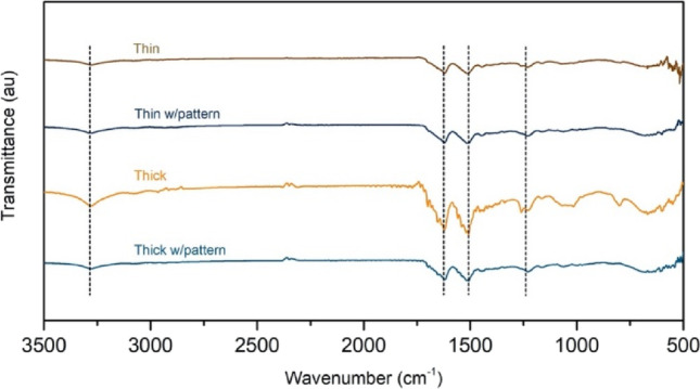
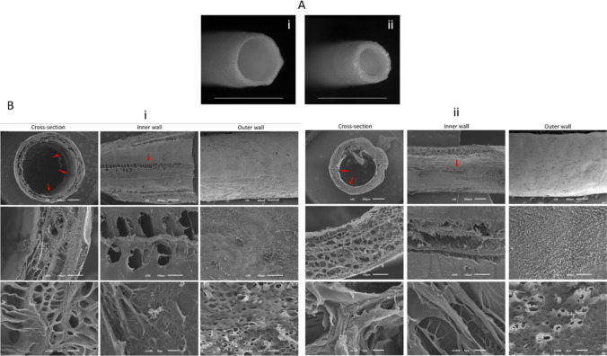
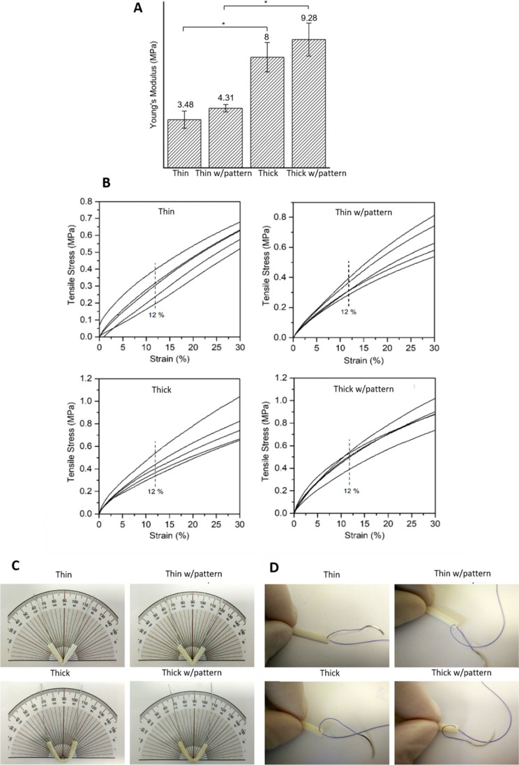
Peripheral nerve injuries represent a major clinical challenge, if nerve ends retract, there is no spontaneous regeneration, and grafts are required to proximate the nerve ends and give continuity to the nerve. The nerve guidance conduits (NGCs) presented in this work are silk fibroin (SF)-based, which is biocompatible and very versatile. The formation of conduits is obtained by forming a covalently cross-linked hydrogel in two concentric moulds, and the inner longitudinally aligned pattern of the SF NGCs is obtained through the use of a patterned inner mould. SF NGCs with two wall thicknesses of ~ 200 to ~ 400 μm are synthesized. Their physicochemical and mechanical characteristics have shown improved properties when the wall thickness is thicker such as resistance to kinking, which is of special importance as conduits might also be used to substitute nerves in flexible body parts. The Young modulus is higher for conduits with inner pattern, and none of the conduits has shown any salt deposition in presence of simulated body fluid, meaning they do not calcify; thus, the regeneration does not get impaired when conduits have contact with body fluids. In vitro studies demonstrated the biocompatibility of the SF NGCs; proliferation is enhanced when iSCs are cultured on top of conduits with longitudinally aligned pattern. BJ fibroblasts cannot infiltrate through the SF wall, avoiding scar tissue formation on the lumen of the graft when used in vivo. These conduits have been demonstrated to be very versatile and fulfil with the requirements for their use in PNR.
Supplementary information: The online version contains supplementary material available at 10.1007/s44164-023-00050-3.


 分享
分享

 求助内容:
求助内容: 应助结果提醒方式:
 扫码关注我们
