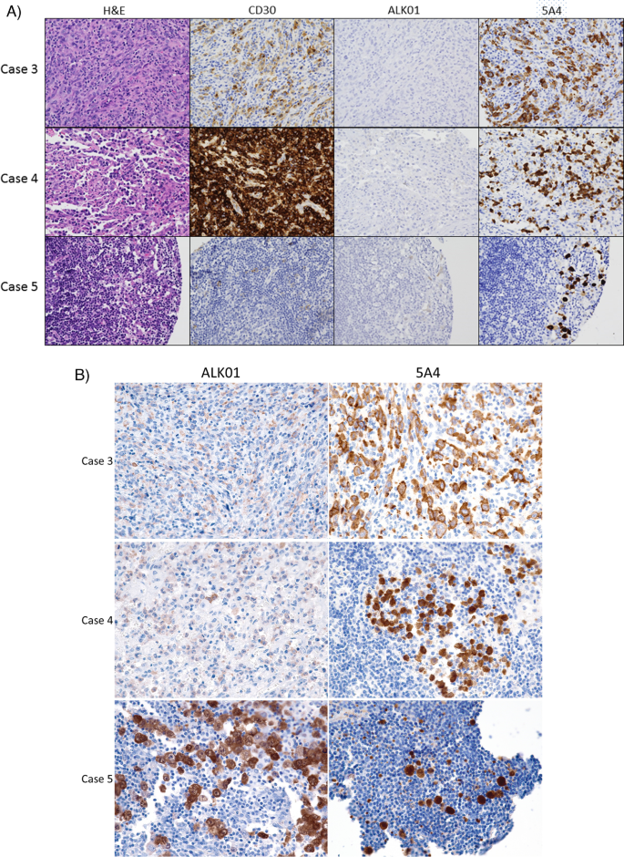
Comparison of two immunohistochemical staining protocols for ALK demonstrates non-inferiority of a 5A4 clone-based protocol versus an ALK01 clone-based protocol for the diagnosis of ALK + anaplastic large cell lymphoma.
Sebastian Fernandez-Pol, Cristiane R Ferreira, Vidhya Manohar, José Antonio Sanches, Luis A P C Lage, Juliana Pereira, Maria C N Zerbini, Dita Gratzinger, Yasodha Natkunam
{"title":"Comparison of two immunohistochemical staining protocols for ALK demonstrates non-inferiority of a 5A4 clone-based protocol versus an ALK01 clone-based protocol for the diagnosis of ALK + anaplastic large cell lymphoma.","authors":"Sebastian Fernandez-Pol, Cristiane R Ferreira, Vidhya Manohar, José Antonio Sanches, Luis A P C Lage, Juliana Pereira, Maria C N Zerbini, Dita Gratzinger, Yasodha Natkunam","doi":"10.1007/s12308-023-00531-0","DOIUrl":null,"url":null,"abstract":"<p><p>Detection of ALK rearrangement and/or expression of the ALK protein is an essential component in the evaluation of many neoplasms. Variability has been reported in the ability of different antibody clones to detect ALK expression. The ALK01 clone is commonly used to detect ALK expression in ALK-positive anaplastic large cell lymphoma (ALK + ALCL). However, this clone has been shown to lack sensitivity when used for solid tumors. The aim of this study was to determine if our high-sensitivity 5A4-based immunohistochemistry protocol is non-inferior to our ALK01-based protocol for the detection of ALK expression in ALK + ALCL. To compare the two protocols, we stained tissue microarrays of 126 hematolymphoid neoplasms and an additional 21 primary cutaneous ALK-negative anaplastic large cell lymphomas with both protocols. All 28 ALK + ALCL samples that were positive for the ALK01 antibody were also positive for the 5A4 clone. Three cases on the tissue microarray that were negative with the ALK01 antibody were clearly positive with the 5A4 antibody. We subsequently stained whole tissue sections of these three cases with the ALK01 antibody and found that these three cases were indeed positive with the ALK01 protocol, suggesting that the absence of staining on the tissue microarray samples was due to a combination of sampling error as well as a dimmer signal with the ALK01 protocol. Our study demonstrates that our 5A4-based protocol is non-inferior to the ALK01 antibody for the diagnosis of ALK-positive anaplastic large cell lymphoma, thus allowing our laboratory to discontinue the use of the ALK01-based protocol.</p>","PeriodicalId":47101,"journal":{"name":"CYBERNETICS AND SYSTEMS ANALYSIS","volume":"38 1","pages":"1-5"},"PeriodicalIF":0.6000,"publicationDate":"2023-03-01","publicationTypes":"Journal Article","fieldsOfStudy":null,"isOpenAccess":false,"openAccessPdf":"https://www.ncbi.nlm.nih.gov/pmc/articles/PMC10766797/pdf/","citationCount":"0","resultStr":null,"platform":"Semanticscholar","paperid":null,"PeriodicalName":"CYBERNETICS AND SYSTEMS ANALYSIS","FirstCategoryId":"1085","ListUrlMain":"https://doi.org/10.1007/s12308-023-00531-0","RegionNum":0,"RegionCategory":null,"ArticlePicture":[],"TitleCN":null,"AbstractTextCN":null,"PMCID":null,"EPubDate":"2023/1/31 0:00:00","PubModel":"Epub","JCR":"Q4","JCRName":"MATHEMATICS, INTERDISCIPLINARY APPLICATIONS","Score":null,"Total":0}
引用次数: 0
Abstract
Detection of ALK rearrangement and/or expression of the ALK protein is an essential component in the evaluation of many neoplasms. Variability has been reported in the ability of different antibody clones to detect ALK expression. The ALK01 clone is commonly used to detect ALK expression in ALK-positive anaplastic large cell lymphoma (ALK + ALCL). However, this clone has been shown to lack sensitivity when used for solid tumors. The aim of this study was to determine if our high-sensitivity 5A4-based immunohistochemistry protocol is non-inferior to our ALK01-based protocol for the detection of ALK expression in ALK + ALCL. To compare the two protocols, we stained tissue microarrays of 126 hematolymphoid neoplasms and an additional 21 primary cutaneous ALK-negative anaplastic large cell lymphomas with both protocols. All 28 ALK + ALCL samples that were positive for the ALK01 antibody were also positive for the 5A4 clone. Three cases on the tissue microarray that were negative with the ALK01 antibody were clearly positive with the 5A4 antibody. We subsequently stained whole tissue sections of these three cases with the ALK01 antibody and found that these three cases were indeed positive with the ALK01 protocol, suggesting that the absence of staining on the tissue microarray samples was due to a combination of sampling error as well as a dimmer signal with the ALK01 protocol. Our study demonstrates that our 5A4-based protocol is non-inferior to the ALK01 antibody for the diagnosis of ALK-positive anaplastic large cell lymphoma, thus allowing our laboratory to discontinue the use of the ALK01-based protocol.
期刊介绍:
Cybernetics and System Analysis publishes articles on: software and hardware;algorithm theory and languages;programming and programming theory;optimization; operations research;digital and analog methods;hybrid systems;machine-machine and man-machine interfacing. Simulation, pattern recognition, artificial intelligence, finite automata, switching theory, and computer logic are also covered. The journal focuses on fresh formulations of problems and new methods of investigation.Cybernetics and System Analysis is a translation of the Ukrainian journal Kibernetika i Sistemnyi Analiz. The Russian Volume Year is published in English from April. All articles are peer reviewed.


 分享
分享
 求助内容:
求助内容: 应助结果提醒方式:
 扫码关注我们
