{"title":"Mouse vaginal development with lateral enlargement at late embryonic stages and caudal elongation after birth","authors":"Masayo Harada, Keiichi Akita","doi":"10.1111/cga.12502","DOIUrl":null,"url":null,"abstract":"<p>Müllerian ducts give rise to the oviducts, uterus, cervix, and vagina. During female reproductive tract development in mice, the bilateral Müllerian duct epithelium grows caudally until reaching the urogenital sinus epithelium. This is followed by further caudal growth with the reduction of the urogenital sinus epithelium. Finally, the vaginal epithelium of adult mice is entirely derived from the Müllerian duct epithelium. Here, we explored the mechanisms underlying mouse vaginal development via cell proliferation, apoptosis, and lineage analyses. We found that at the late embryonic stages, apoptosis occurred at the attachment site of bilateral Müllerian duct epithelia below the cervix, resulting in bilateral lumen traffic. The Müllerian duct epithelium was enclosed by the urogenital sinus epithelium at their boundary region on embryonic day (E) 16.5, whereas the Müllerian duct epithelium encased the urogenital sinus epithelium at postnatal day (P) 0 through lateral enlargement. Lateral Müllerian duct enlargement was accompanied by focal ERK activation within the curved epithelial tips and the specific localization of mitotic nuclei on the luminal side of the Müllerian duct epithelial layer at E17.5. Descent of the Müllerian duct epithelium and shortening of the urogenital sinus epithelium occurred rapidly after birth, accompanied by cell proliferation in the Müllerian duct epithelium and its peripheral mesenchymal tissues as well as intense apoptosis in the urogenital sinus epithelium around their boundary region. Urogenital sinus epithelium was localized at the base of the vagina at P7. In conclusion, the mouse vagina develops laterally at the late embryonic stages and caudally after birth.</p>","PeriodicalId":10626,"journal":{"name":"Congenital Anomalies","volume":"63 2","pages":"30-39"},"PeriodicalIF":1.6000,"publicationDate":"2022-12-14","publicationTypes":"Journal Article","fieldsOfStudy":null,"isOpenAccess":false,"openAccessPdf":"https://onlinelibrary.wiley.com/doi/epdf/10.1111/cga.12502","citationCount":"0","resultStr":null,"platform":"Semanticscholar","paperid":null,"PeriodicalName":"Congenital Anomalies","FirstCategoryId":"3","ListUrlMain":"https://onlinelibrary.wiley.com/doi/10.1111/cga.12502","RegionNum":4,"RegionCategory":"医学","ArticlePicture":[],"TitleCN":null,"AbstractTextCN":null,"PMCID":null,"EPubDate":"","PubModel":"","JCR":"Q3","JCRName":"PEDIATRICS","Score":null,"Total":0}
引用次数: 0
Abstract
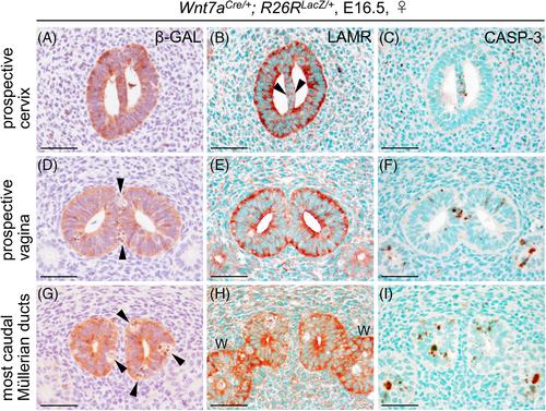
Müllerian ducts give rise to the oviducts, uterus, cervix, and vagina. During female reproductive tract development in mice, the bilateral Müllerian duct epithelium grows caudally until reaching the urogenital sinus epithelium. This is followed by further caudal growth with the reduction of the urogenital sinus epithelium. Finally, the vaginal epithelium of adult mice is entirely derived from the Müllerian duct epithelium. Here, we explored the mechanisms underlying mouse vaginal development via cell proliferation, apoptosis, and lineage analyses. We found that at the late embryonic stages, apoptosis occurred at the attachment site of bilateral Müllerian duct epithelia below the cervix, resulting in bilateral lumen traffic. The Müllerian duct epithelium was enclosed by the urogenital sinus epithelium at their boundary region on embryonic day (E) 16.5, whereas the Müllerian duct epithelium encased the urogenital sinus epithelium at postnatal day (P) 0 through lateral enlargement. Lateral Müllerian duct enlargement was accompanied by focal ERK activation within the curved epithelial tips and the specific localization of mitotic nuclei on the luminal side of the Müllerian duct epithelial layer at E17.5. Descent of the Müllerian duct epithelium and shortening of the urogenital sinus epithelium occurred rapidly after birth, accompanied by cell proliferation in the Müllerian duct epithelium and its peripheral mesenchymal tissues as well as intense apoptosis in the urogenital sinus epithelium around their boundary region. Urogenital sinus epithelium was localized at the base of the vagina at P7. In conclusion, the mouse vagina develops laterally at the late embryonic stages and caudally after birth.
期刊介绍:
Congenital Anomalies is the official English language journal of the Japanese Teratology Society, and publishes original articles in laboratory as well as clinical research in all areas of abnormal development and related fields, from all over the world. Although contributions by members of the teratology societies affiliated with The International Federation of Teratology Societies are given priority, contributions from non-members are welcomed.


 分享
分享
 求助内容:
求助内容: 应助结果提醒方式:
 扫码关注我们
