Abraham Castro-Cruz, Olga M. Echeverría, Luis Sánchez-Sánchez, Israel Muñoz-Velasco, Silvia Juárez-Chavero, Nayeli Torres-Ramírez, Gerardo H. Vázquez-Nin, María Luisa Escobar
{"title":"Dissection of the autophagic route in oocytes from atretic follicles","authors":"Abraham Castro-Cruz, Olga M. Echeverría, Luis Sánchez-Sánchez, Israel Muñoz-Velasco, Silvia Juárez-Chavero, Nayeli Torres-Ramírez, Gerardo H. Vázquez-Nin, María Luisa Escobar","doi":"10.1111/boc.202200046","DOIUrl":null,"url":null,"abstract":"<div>\n \n \n <section>\n \n <h3> Background Information</h3>\n \n <p>Autophagy is a conserved process that functions as a cytoprotective mechanism; it may function as a cell death process called programmed cell death type II. There is considerable evidence for the presence of autophagic cell death during oocyte elimination in prepubertal rats. However, the mechanisms involved in this process have not been deciphered.</p>\n </section>\n \n <section>\n \n <h3> Results</h3>\n \n <p>Our observations revealed autophagic cell death in oocytes with increased labeling of the autophagic proteins Beclin 1, light chain 3 A (LC3 A), and lysosomal-associated membrane protein 1 (Lamp1). Furthermore, mTOR and phosphorylated (p)-mTOR (S2448) proteins were significantly decreased in oocytes with increased levels of autophagic proteins, indicating autophagic activation. Moreover, phosphorylated protein kinase B (p-AKT) was not expressed by oocytes, but mitogen-activated protein kinase/extracellular signalregulated kinase (MAPK/ERK) signaling was observed. Additionally, selective and elevated mitochondrial degradation was identified in altered oocytes.</p>\n </section>\n \n <section>\n \n <h3> Conclusions</h3>\n \n <p>All these results suggest that mTOR downregulation, which promotes autophagy, could be mediated by low energy levels and sustained starvation involving the phosphoinositide 3-kinase (PI3K)/AKT/mTOR and MAPK/ERK pathways.</p>\n </section>\n \n <section>\n \n <h3> Significance</h3>\n \n <p>In this work, we analyzed the manner in which autophagy is carried out in oocytes undergoing autophagic cell death by studying the behavior of proteins involved in different steps of the autophagic pathway.</p>\n </section>\n </div>","PeriodicalId":8859,"journal":{"name":"Biology of the Cell","volume":"115 3","pages":""},"PeriodicalIF":2.4000,"publicationDate":"2022-12-26","publicationTypes":"Journal Article","fieldsOfStudy":null,"isOpenAccess":false,"openAccessPdf":"https://onlinelibrary.wiley.com/doi/epdf/10.1111/boc.202200046","citationCount":"0","resultStr":null,"platform":"Semanticscholar","paperid":null,"PeriodicalName":"Biology of the Cell","FirstCategoryId":"99","ListUrlMain":"https://onlinelibrary.wiley.com/doi/10.1111/boc.202200046","RegionNum":4,"RegionCategory":"生物学","ArticlePicture":[],"TitleCN":null,"AbstractTextCN":null,"PMCID":null,"EPubDate":"","PubModel":"","JCR":"Q4","JCRName":"CELL BIOLOGY","Score":null,"Total":0}
引用次数: 0
Abstract
Background Information
Autophagy is a conserved process that functions as a cytoprotective mechanism; it may function as a cell death process called programmed cell death type II. There is considerable evidence for the presence of autophagic cell death during oocyte elimination in prepubertal rats. However, the mechanisms involved in this process have not been deciphered.
Results
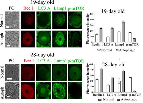
Our observations revealed autophagic cell death in oocytes with increased labeling of the autophagic proteins Beclin 1, light chain 3 A (LC3 A), and lysosomal-associated membrane protein 1 (Lamp1). Furthermore, mTOR and phosphorylated (p)-mTOR (S2448) proteins were significantly decreased in oocytes with increased levels of autophagic proteins, indicating autophagic activation. Moreover, phosphorylated protein kinase B (p-AKT) was not expressed by oocytes, but mitogen-activated protein kinase/extracellular signalregulated kinase (MAPK/ERK) signaling was observed. Additionally, selective and elevated mitochondrial degradation was identified in altered oocytes.
Conclusions
All these results suggest that mTOR downregulation, which promotes autophagy, could be mediated by low energy levels and sustained starvation involving the phosphoinositide 3-kinase (PI3K)/AKT/mTOR and MAPK/ERK pathways.
Significance
In this work, we analyzed the manner in which autophagy is carried out in oocytes undergoing autophagic cell death by studying the behavior of proteins involved in different steps of the autophagic pathway.
期刊介绍:
The journal publishes original research articles and reviews on all aspects of cellular, molecular and structural biology, developmental biology, cell physiology and evolution. It will publish articles or reviews contributing to the understanding of the elementary biochemical and biophysical principles of live matter organization from the molecular, cellular and tissues scales and organisms.
This includes contributions directed towards understanding biochemical and biophysical mechanisms, structure-function relationships with respect to basic cell and tissue functions, development, development/evolution relationship, morphogenesis, stem cell biology, cell biology of disease, plant cell biology, as well as contributions directed toward understanding integrated processes at the organelles, cell and tissue levels. Contributions using approaches such as high resolution imaging, live imaging, quantitative cell biology and integrated biology; as well as those using innovative genetic and epigenetic technologies, ex-vivo tissue engineering, cellular, tissue and integrated functional analysis, and quantitative biology and modeling to demonstrate original biological principles are encouraged.


 分享
分享
 求助内容:
求助内容: 应助结果提醒方式:
 扫码关注我们
