S M Bennji, A H Al-Kindi, B Jayakrishnan, A Al Shehhi, B Itkhan
{"title":"Cheerios sign: A rare sign on chest computed tomography.","authors":"S M Bennji, A H Al-Kindi, B Jayakrishnan, A Al Shehhi, B Itkhan","doi":"10.7196/AJTCCM.2022.v28i4.251","DOIUrl":null,"url":null,"abstract":"A 44-year-old man initially presented to a tertiary hospital in Oman in August 2020 with a right iliopsoas mass, which was found to be a high-grade myxofibrosarcoma. At initial presentation, there was no evidence of metastases anywhere. He underwent local resection with free surgical margins followed by adjuvant radiotherapy. A year later, he presented with mild haemoptysis for a few weeks with no other constitutional symptoms. High-resolution computed tomography (CT) showed a nodule with a central lucent cavity and a surrounding ground-glass rim known as the Cheerios sign in the right lower lobe (Fig. 1A). Bronchoscopy revealed spots of fresh blood coming from the lateral basal segment of the right lower lobe. Bronchoalveolar lavage revealed no malignant cells on cytology, no growth in bacterial and fungal cultures and a negative GeneXpert test. The patient underwent video-assisted thoracoscopic surgery and wedge resection of the nodule. Histopathological findings confirmed metastatic myxofibrosarcoma (Fig. 2). Subsequently, the patient was subjected to adjuvant chemotherapy. Cheerios sign is a rare finding on CT, defined as a nodule with central radiolucency, resembling the ring-shaped Cheerios breakfast cereal (Fig 1B).[1] It was described for the first time by Reed and O’Neil in 1993.[2] It presents as a small ring-like uniform shadowing embedded in the normal surrounding lung. It is formed by peribronchiolar proliferation of non-malignant or malignant cells as in this case.[3,4] The most common causes of cheerios sign are pulmonary Langerhans cell histiocytosis and lepidic growth of pulmonary adenocarcinoma. Differential diagnoses include granulomatosis with polyangitis, rheumatoid nodules or fungal and mycobacterial infections.[1] Although cavitary metastasis is seen in sarcomas, it has not been reported in myxofibrosarcomas. Cheerios sign in patients with underlying malignancy should be considered metastatic until proven otherwise. Cheerios sign: A rare sign on chest computed tomography","PeriodicalId":52847,"journal":{"name":"African Journal of Thoracic and Critical Care Medicine","volume":"28 4","pages":""},"PeriodicalIF":0.0000,"publicationDate":"2022-01-01","publicationTypes":"Journal Article","fieldsOfStudy":null,"isOpenAccess":false,"openAccessPdf":"https://ftp.ncbi.nlm.nih.gov/pub/pmc/oa_pdf/e9/55/AJTCCM-28-4-251.PMC9979626.pdf","citationCount":"6","resultStr":null,"platform":"Semanticscholar","paperid":null,"PeriodicalName":"African Journal of Thoracic and Critical Care Medicine","FirstCategoryId":"1085","ListUrlMain":"https://doi.org/10.7196/AJTCCM.2022.v28i4.251","RegionNum":0,"RegionCategory":null,"ArticlePicture":[],"TitleCN":null,"AbstractTextCN":null,"PMCID":null,"EPubDate":"","PubModel":"","JCR":"Q3","JCRName":"Medicine","Score":null,"Total":0}
引用次数: 6
Abstract
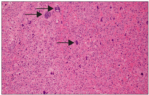
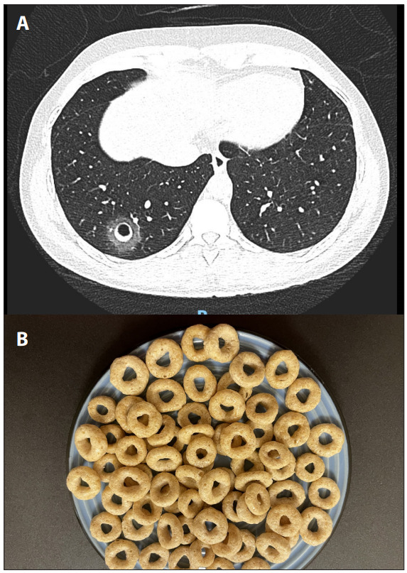
A 44-year-old man initially presented to a tertiary hospital in Oman in August 2020 with a right iliopsoas mass, which was found to be a high-grade myxofibrosarcoma. At initial presentation, there was no evidence of metastases anywhere. He underwent local resection with free surgical margins followed by adjuvant radiotherapy. A year later, he presented with mild haemoptysis for a few weeks with no other constitutional symptoms. High-resolution computed tomography (CT) showed a nodule with a central lucent cavity and a surrounding ground-glass rim known as the Cheerios sign in the right lower lobe (Fig. 1A). Bronchoscopy revealed spots of fresh blood coming from the lateral basal segment of the right lower lobe. Bronchoalveolar lavage revealed no malignant cells on cytology, no growth in bacterial and fungal cultures and a negative GeneXpert test. The patient underwent video-assisted thoracoscopic surgery and wedge resection of the nodule. Histopathological findings confirmed metastatic myxofibrosarcoma (Fig. 2). Subsequently, the patient was subjected to adjuvant chemotherapy. Cheerios sign is a rare finding on CT, defined as a nodule with central radiolucency, resembling the ring-shaped Cheerios breakfast cereal (Fig 1B).[1] It was described for the first time by Reed and O’Neil in 1993.[2] It presents as a small ring-like uniform shadowing embedded in the normal surrounding lung. It is formed by peribronchiolar proliferation of non-malignant or malignant cells as in this case.[3,4] The most common causes of cheerios sign are pulmonary Langerhans cell histiocytosis and lepidic growth of pulmonary adenocarcinoma. Differential diagnoses include granulomatosis with polyangitis, rheumatoid nodules or fungal and mycobacterial infections.[1] Although cavitary metastasis is seen in sarcomas, it has not been reported in myxofibrosarcomas. Cheerios sign in patients with underlying malignancy should be considered metastatic until proven otherwise. Cheerios sign: A rare sign on chest computed tomography


 分享
分享
 求助内容:
求助内容: 应助结果提醒方式:
 扫码关注我们
