Ayoung Kang, Sung Hwan Cho, Byung-Soo Park, Gyung Mo Son, Hyun Sung Kim, Jae-Joon Kim, Su Jin Kim, Dong Hoon Shin, Tae Un Kim
{"title":"Perianal extragastrointestinal stromal tumor.","authors":"Ayoung Kang, Sung Hwan Cho, Byung-Soo Park, Gyung Mo Son, Hyun Sung Kim, Jae-Joon Kim, Su Jin Kim, Dong Hoon Shin, Tae Un Kim","doi":"10.14216/kjco.20021","DOIUrl":null,"url":null,"abstract":"<p><p>An extragastrointestinal stromal tumor (EGIST) is a gastrointestinal stromal tumor that arises outside of the gastrointestinal tract. Most EGISTs are located in the omentum, mesentery, and retroperitoneum. The occurrence of an EGIST at the perianal region is very rare. Herein, we report our experience with EGISTs in the perianal area and review the literature. A 70-year-old man presented to our hospital with a 2-year history of anal discomfort. A pelvic magnetic resonance imaging scan showed a homogenous, well-defined, soft tissue density mass. The patient underwent mass excision, and the pathological examination confirmed that the mass was an EGIST. The size of the tumor was 4.3×3.2 cm, and the mitotic count was 1 per 50 high-power fields. The tumor cells were immunohistochemically positive for KIT and CD34 but were negative for S-100 and alpha-smooth muscle actin. There were no other abnormal findings in the gastrointestinal tract; upon pathological review, this case was confirmed as perianal EGIST. Therefore, EGIST should be considered as a differential diagnosis of perianal masses.</p>","PeriodicalId":74045,"journal":{"name":"Korean journal of clinical oncology","volume":"16 2","pages":"138-141"},"PeriodicalIF":0.0000,"publicationDate":"2020-12-01","publicationTypes":"Journal Article","fieldsOfStudy":null,"isOpenAccess":false,"openAccessPdf":"https://ftp.ncbi.nlm.nih.gov/pub/pmc/oa_pdf/24/27/kjco-16-2-138.PMC9942734.pdf","citationCount":"0","resultStr":null,"platform":"Semanticscholar","paperid":null,"PeriodicalName":"Korean journal of clinical oncology","FirstCategoryId":"1085","ListUrlMain":"https://doi.org/10.14216/kjco.20021","RegionNum":0,"RegionCategory":null,"ArticlePicture":[],"TitleCN":null,"AbstractTextCN":null,"PMCID":null,"EPubDate":"","PubModel":"","JCR":"","JCRName":"","Score":null,"Total":0}
引用次数: 0
Abstract
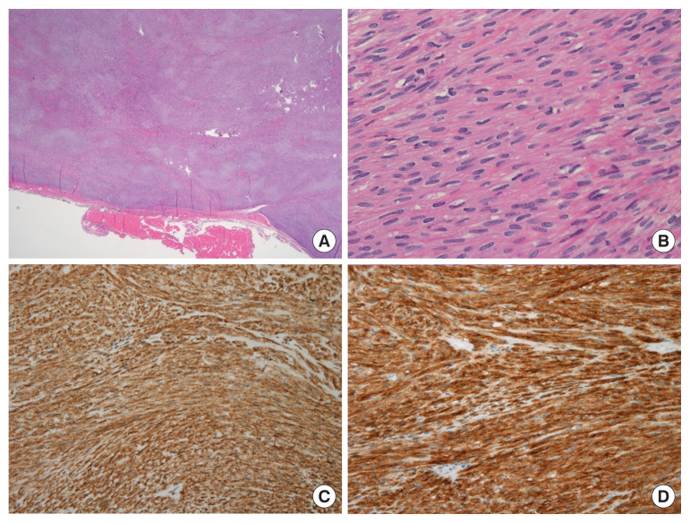
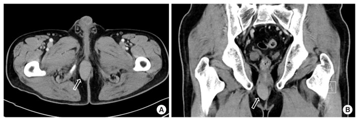
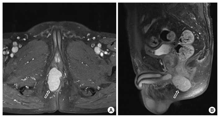
An extragastrointestinal stromal tumor (EGIST) is a gastrointestinal stromal tumor that arises outside of the gastrointestinal tract. Most EGISTs are located in the omentum, mesentery, and retroperitoneum. The occurrence of an EGIST at the perianal region is very rare. Herein, we report our experience with EGISTs in the perianal area and review the literature. A 70-year-old man presented to our hospital with a 2-year history of anal discomfort. A pelvic magnetic resonance imaging scan showed a homogenous, well-defined, soft tissue density mass. The patient underwent mass excision, and the pathological examination confirmed that the mass was an EGIST. The size of the tumor was 4.3×3.2 cm, and the mitotic count was 1 per 50 high-power fields. The tumor cells were immunohistochemically positive for KIT and CD34 but were negative for S-100 and alpha-smooth muscle actin. There were no other abnormal findings in the gastrointestinal tract; upon pathological review, this case was confirmed as perianal EGIST. Therefore, EGIST should be considered as a differential diagnosis of perianal masses.


 分享
分享

 求助内容:
求助内容: 应助结果提醒方式:
 扫码关注我们
