Diagnostic efficacy of diffusion-weighted imaging and semiquantitative and quantitative dynamic contrast-enhanced magnetic resonance imaging in salivary gland tumors.
{"title":"Diagnostic efficacy of diffusion-weighted imaging and semiquantitative and quantitative dynamic contrast-enhanced magnetic resonance imaging in salivary gland tumors.","authors":"Erkan Gökçe, Murat Beyhan","doi":"10.4329/wjr.v15.i1.20","DOIUrl":null,"url":null,"abstract":"<p><strong>Background: </strong>Increased use of functional magnetic resonance imaging (MRI) methods such as diffusion-weighted imaging (DWI) and dynamic contrast-enhanced (DCE) MRI consisting of sequential contrast series, allows us to obtain more information on the microstructure, cellularity, interstitial distance, and vascularity of tumors, which has increased the discrimination power for benign and malignant salivary gland tumors (SGTs). In the last few years, quantitative DCE MRI data containing T1 perfusion parameters (K<sub>trans</sub>, K<sub>ep</sub> and V<sub>e</sub>), were reported to contribute to the differentiation of benign or malignant subtypes in SGTs.</p><p><strong>Aim: </strong>To evaluate the diagnostic efficacy of DWI and semiquantitative and quantitative perfusion MRI parameters in SGTs.</p><p><strong>Methods: </strong>Diffusion MRI [apparent diffusion coefficient (ADC) value] with a 1.5 T MR machine, semiquantitative perfusion MRI [time intensity curve (TIC) pattern], and quantitative perfusion MRI examinations (K<sub>trans</sub>, K<sub>ep</sub> and V<sub>e</sub>) of 73 tumors in 67 patients with histopathological diagnosis performed from 2017 to 2021 were retrospectively evaluated. In the ADC value and semiquantitative perfusion MRI measurements, cystic components of the tumors were not considered, and the region of interest (ROI) was manually placed through the widest axial section of the tumor. TIC patterns were divided into four groups: Type A = T<sub>peak</sub> > 120 s; type B = T<sub>peak</sub> ≤ 120 s, washout ratio (WR) ≥ 30%; type C = T<sub>peak</sub> ≤ 120 s, WR < 30%; and type D = flat TIC. For the quantitative perfusion MRI analysis, a 3D ROI was placed in the largest solid component of the tumor, and the K<sub>trans</sub>, K<sub>ep</sub> and V<sub>e</sub> values were automatically generated.</p><p><strong>Results: </strong>The majority of SGTs were located in the parotid glands (86.3%). Of all the SGTs, 68.5% were benign and 31.5% were malignant. Significant differences were found for ADC values among pleomorphic adenomas (PMAs), Warthin's tumors (WTs), and malignant tumors (MTs) (<i>P</i> < 0.001). PMAs had type A and WTs had type B TIC pattern while the vast majority of MTs and other benign tumors (OBTs) (54.5% and 45.5%, respectively) displayed type C TIC pattern. PMAs showed no washout, while the highest mean WR was observed in WTs (59% ± 11%). K<sub>trans</sub> values of PMAs, WTs, OBTs, and MTs were not significantly different. K<sub>ep</sub> values of PMAs and WTs were significantly different from those of OBTs and MTs. Mean V<sub>e</sub> value of WTs was significantly different from those of PMAs, OBTs, and MTs (<i>P</i> < 0.001).</p><p><strong>Conclusion: </strong>The use of quantitative DCE parameters along with diffusion MRI and semiquantitative contrast-enhanced MRI in SGTs could improve the diagnostic accuracy.</p>","PeriodicalId":23819,"journal":{"name":"World journal of radiology","volume":"15 1","pages":"20-31"},"PeriodicalIF":1.5000,"publicationDate":"2023-01-28","publicationTypes":"Journal Article","fieldsOfStudy":null,"isOpenAccess":false,"openAccessPdf":"https://ftp.ncbi.nlm.nih.gov/pub/pmc/oa_pdf/a3/ad/WJR-15-20.PMC9884336.pdf","citationCount":"0","resultStr":null,"platform":"Semanticscholar","paperid":null,"PeriodicalName":"World journal of radiology","FirstCategoryId":"1085","ListUrlMain":"https://doi.org/10.4329/wjr.v15.i1.20","RegionNum":0,"RegionCategory":null,"ArticlePicture":[],"TitleCN":null,"AbstractTextCN":null,"PMCID":null,"EPubDate":"","PubModel":"","JCR":"Q3","JCRName":"RADIOLOGY, NUCLEAR MEDICINE & MEDICAL IMAGING","Score":null,"Total":0}
引用次数: 0
Abstract
Background: Increased use of functional magnetic resonance imaging (MRI) methods such as diffusion-weighted imaging (DWI) and dynamic contrast-enhanced (DCE) MRI consisting of sequential contrast series, allows us to obtain more information on the microstructure, cellularity, interstitial distance, and vascularity of tumors, which has increased the discrimination power for benign and malignant salivary gland tumors (SGTs). In the last few years, quantitative DCE MRI data containing T1 perfusion parameters (Ktrans, Kep and Ve), were reported to contribute to the differentiation of benign or malignant subtypes in SGTs.
Aim: To evaluate the diagnostic efficacy of DWI and semiquantitative and quantitative perfusion MRI parameters in SGTs.
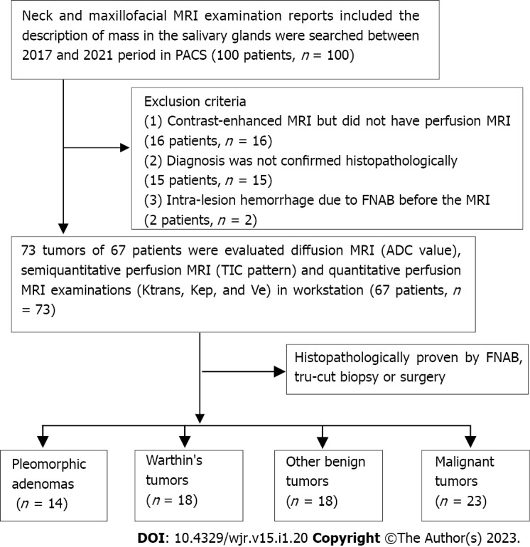
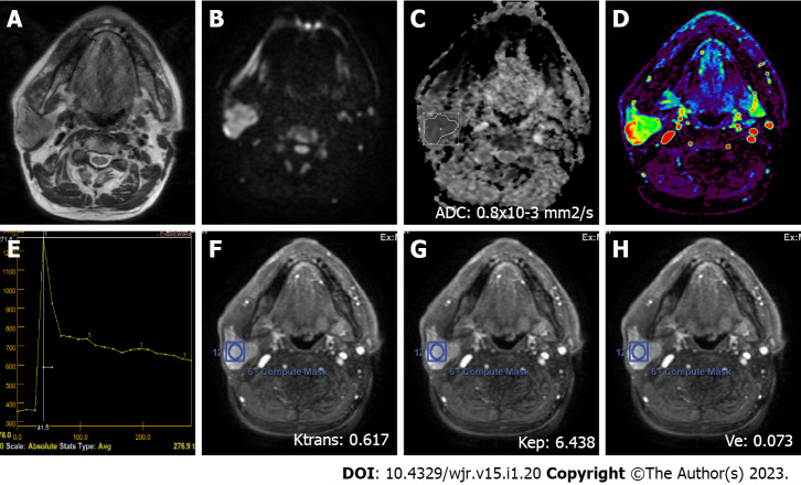
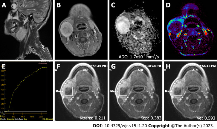
Methods: Diffusion MRI [apparent diffusion coefficient (ADC) value] with a 1.5 T MR machine, semiquantitative perfusion MRI [time intensity curve (TIC) pattern], and quantitative perfusion MRI examinations (Ktrans, Kep and Ve) of 73 tumors in 67 patients with histopathological diagnosis performed from 2017 to 2021 were retrospectively evaluated. In the ADC value and semiquantitative perfusion MRI measurements, cystic components of the tumors were not considered, and the region of interest (ROI) was manually placed through the widest axial section of the tumor. TIC patterns were divided into four groups: Type A = Tpeak > 120 s; type B = Tpeak ≤ 120 s, washout ratio (WR) ≥ 30%; type C = Tpeak ≤ 120 s, WR < 30%; and type D = flat TIC. For the quantitative perfusion MRI analysis, a 3D ROI was placed in the largest solid component of the tumor, and the Ktrans, Kep and Ve values were automatically generated.
Results: The majority of SGTs were located in the parotid glands (86.3%). Of all the SGTs, 68.5% were benign and 31.5% were malignant. Significant differences were found for ADC values among pleomorphic adenomas (PMAs), Warthin's tumors (WTs), and malignant tumors (MTs) (P < 0.001). PMAs had type A and WTs had type B TIC pattern while the vast majority of MTs and other benign tumors (OBTs) (54.5% and 45.5%, respectively) displayed type C TIC pattern. PMAs showed no washout, while the highest mean WR was observed in WTs (59% ± 11%). Ktrans values of PMAs, WTs, OBTs, and MTs were not significantly different. Kep values of PMAs and WTs were significantly different from those of OBTs and MTs. Mean Ve value of WTs was significantly different from those of PMAs, OBTs, and MTs (P < 0.001).
Conclusion: The use of quantitative DCE parameters along with diffusion MRI and semiquantitative contrast-enhanced MRI in SGTs could improve the diagnostic accuracy.


 分享
分享
 求助内容:
求助内容: 应助结果提醒方式:
 扫码关注我们
