{"title":"Pre-treatment with IL-6 potentiates β-cell death induced by pro-inflammatory cytokines.","authors":"V R Oliveira, C C Paula, S Taniguchi, F Ortis","doi":"10.1186/s12860-023-00476-3","DOIUrl":null,"url":null,"abstract":"<p><strong>Background: </strong>Type I Diabetes mellitus (T1D) is characterized by a specific destruction of β-cells by the immune system. During this process pro-inflammatory cytokines are released in the pancreatic islets and contribute for β-cells demise. Cytokine-induced iNOS activation, via NF-κB, is implicated in induction of β-cells death, which includes ER stress activation. Physical exercise has been used as an adjunct for better glycemic control in patients with T1D, since it is able to increase glucose uptake independent of insulin. Recently, it was observed that the release of IL-6 by skeletal muscle, during physical exercise, could prevent β-cells death induced by pro-inflammatory cytokines. However, the molecular mechanisms involved in this beneficial effect on β-cells are not yet completely elucidated. Our aim was to evaluate the effect of IL-6 on β-cells exposed to pro-inflammatory cytokines.</p><p><strong>Results: </strong>Pre-treatment with IL-6 sensitized INS-1E cells to cytokine-induced cell death, increasing cytokine-induced iNOS and Caspase-3 expression. Under these conditions, however, there was a decrease in cytokines-induced p-eIF2-α but not p-IRE1expression, proteins related to ER stress. To address if this prevention of adequate UPR response is involved in the increase in β-cells death markers induced by IL-6 pre-treatment, we used a chemical chaperone (TUDCA), which improves ER folding capacity. Use of TUDCA increased cytokines-induced Caspase-3 expression and Bax/Bcl-2 ratio in the presence of IL-6 pre-treatment. However, there is no modulation of p-eIF2-α expression by TUDCA in this condition, with increase of CHOP expression.</p><p><strong>Conclusion: </strong>Treatment with IL-6 alone is not beneficial for β-cells, leading to increased cell death markers and impaired UPR activation. In addition, TUDCA has not been able to restore ER homeostasis or improve β-cells viability under this condition, suggesting that other mechanisms may be involved.</p>","PeriodicalId":9099,"journal":{"name":"BMC Molecular and Cell Biology","volume":"24 1","pages":"11"},"PeriodicalIF":2.7000,"publicationDate":"2023-03-28","publicationTypes":"Journal Article","fieldsOfStudy":null,"isOpenAccess":false,"openAccessPdf":"https://www.ncbi.nlm.nih.gov/pmc/articles/PMC10045109/pdf/","citationCount":"1","resultStr":null,"platform":"Semanticscholar","paperid":null,"PeriodicalName":"BMC Molecular and Cell Biology","FirstCategoryId":"3","ListUrlMain":"https://doi.org/10.1186/s12860-023-00476-3","RegionNum":3,"RegionCategory":"生物学","ArticlePicture":[],"TitleCN":null,"AbstractTextCN":null,"PMCID":null,"EPubDate":"","PubModel":"","JCR":"Q4","JCRName":"CELL BIOLOGY","Score":null,"Total":0}
引用次数: 1
Abstract
Background: Type I Diabetes mellitus (T1D) is characterized by a specific destruction of β-cells by the immune system. During this process pro-inflammatory cytokines are released in the pancreatic islets and contribute for β-cells demise. Cytokine-induced iNOS activation, via NF-κB, is implicated in induction of β-cells death, which includes ER stress activation. Physical exercise has been used as an adjunct for better glycemic control in patients with T1D, since it is able to increase glucose uptake independent of insulin. Recently, it was observed that the release of IL-6 by skeletal muscle, during physical exercise, could prevent β-cells death induced by pro-inflammatory cytokines. However, the molecular mechanisms involved in this beneficial effect on β-cells are not yet completely elucidated. Our aim was to evaluate the effect of IL-6 on β-cells exposed to pro-inflammatory cytokines.
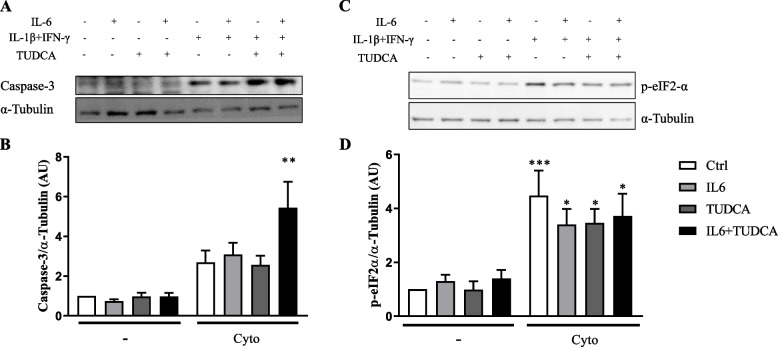
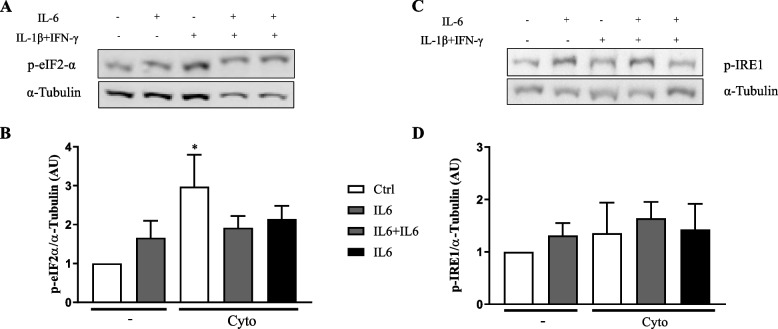
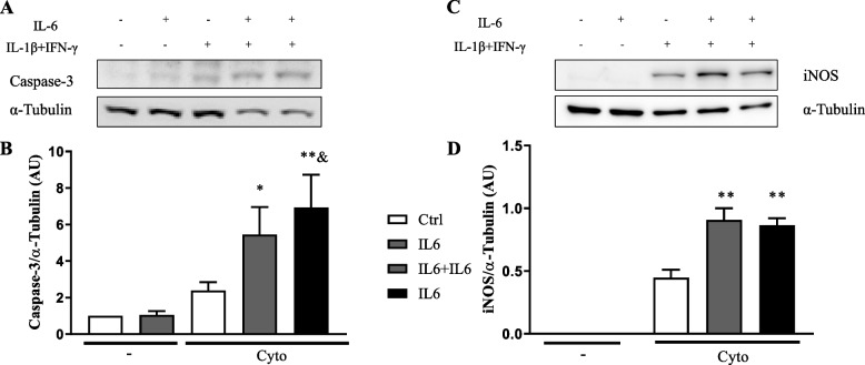
Results: Pre-treatment with IL-6 sensitized INS-1E cells to cytokine-induced cell death, increasing cytokine-induced iNOS and Caspase-3 expression. Under these conditions, however, there was a decrease in cytokines-induced p-eIF2-α but not p-IRE1expression, proteins related to ER stress. To address if this prevention of adequate UPR response is involved in the increase in β-cells death markers induced by IL-6 pre-treatment, we used a chemical chaperone (TUDCA), which improves ER folding capacity. Use of TUDCA increased cytokines-induced Caspase-3 expression and Bax/Bcl-2 ratio in the presence of IL-6 pre-treatment. However, there is no modulation of p-eIF2-α expression by TUDCA in this condition, with increase of CHOP expression.
Conclusion: Treatment with IL-6 alone is not beneficial for β-cells, leading to increased cell death markers and impaired UPR activation. In addition, TUDCA has not been able to restore ER homeostasis or improve β-cells viability under this condition, suggesting that other mechanisms may be involved.


 分享
分享
 求助内容:
求助内容: 应助结果提醒方式:
 扫码关注我们
