David Martínez, Gustavo E Sanchez, Jhonatan Gómez, Luis J Sonda, Luis D Suárez, Carlos S López, Juan J Vega, Daniel A Cepeda
{"title":"Magnetic resonance imaging findings of spontaneous pyomyoma in a premenopausal woman managed with myomectomy: A case report.","authors":"David Martínez, Gustavo E Sanchez, Jhonatan Gómez, Luis J Sonda, Luis D Suárez, Carlos S López, Juan J Vega, Daniel A Cepeda","doi":"10.4329/wjr.v15.i3.83","DOIUrl":null,"url":null,"abstract":"<p><strong>Background: </strong>Acute fibroid complications are rare. However, failure to recognize and treat acute complications expeditiously when they occur can lead to catastrophic, even deadly, complications. Pyomyoma is a rare but potentially fatal condition resulting from infarction and infection of a fibroid through bacterial seeding and direct, hematogenous, or lymphatic dissemination. Even though the diagnosis is established through clinical and laboratory findings, imaging is an important complementary method to support the suspected diagnosis.</p><p><strong>Case summary: </strong>Herein, we report a case of a pyomyoma in a nulliparous woman previously diagnosed with uterine leiomyomatosis according to ultrasound findings. The patient had previously attended the emergency room due to hypogastric pain unresponsive to analgesics. After a week of persistent pain, she developed sepsis without any identifiable foci. Magnetic resonance imaging revealed findings compatible with uterine myomatosis with red degeneration, and a possible diagnosis of a pyomyoma was made according to the imaging findings along with the patient's clinical features. We decided to perform myomectomy (which is an infrequently performed surgical treatment due to the procedure's intrinsic implications) due to the patient's desire to preserve fertility. Histopathologic results revealed a uterine leiomyoma with coagulative and liquefactive necrosis, while the tissue culture showed gram-negative cocci bacteria, which were successfully treated using antibiotic therapy. The patient's health status improved after several days.</p><p><strong>Conclusion: </strong>The main diagnostic tools to evaluate pyomyomas are the clinical and laboratory findings as well as tissue cultures. Nonetheless, magnetic resonance imaging can help to corroborate these findings as well as to better characterize myomas with its different complications.</p>","PeriodicalId":23819,"journal":{"name":"World journal of radiology","volume":"15 3","pages":"83-88"},"PeriodicalIF":1.5000,"publicationDate":"2023-03-28","publicationTypes":"Journal Article","fieldsOfStudy":null,"isOpenAccess":false,"openAccessPdf":"https://ftp.ncbi.nlm.nih.gov/pub/pmc/oa_pdf/1c/c2/WJR-15-83.PMC10080582.pdf","citationCount":"0","resultStr":null,"platform":"Semanticscholar","paperid":null,"PeriodicalName":"World journal of radiology","FirstCategoryId":"1085","ListUrlMain":"https://doi.org/10.4329/wjr.v15.i3.83","RegionNum":0,"RegionCategory":null,"ArticlePicture":[],"TitleCN":null,"AbstractTextCN":null,"PMCID":null,"EPubDate":"","PubModel":"","JCR":"Q3","JCRName":"RADIOLOGY, NUCLEAR MEDICINE & MEDICAL IMAGING","Score":null,"Total":0}
引用次数: 0
Abstract
Background: Acute fibroid complications are rare. However, failure to recognize and treat acute complications expeditiously when they occur can lead to catastrophic, even deadly, complications. Pyomyoma is a rare but potentially fatal condition resulting from infarction and infection of a fibroid through bacterial seeding and direct, hematogenous, or lymphatic dissemination. Even though the diagnosis is established through clinical and laboratory findings, imaging is an important complementary method to support the suspected diagnosis.
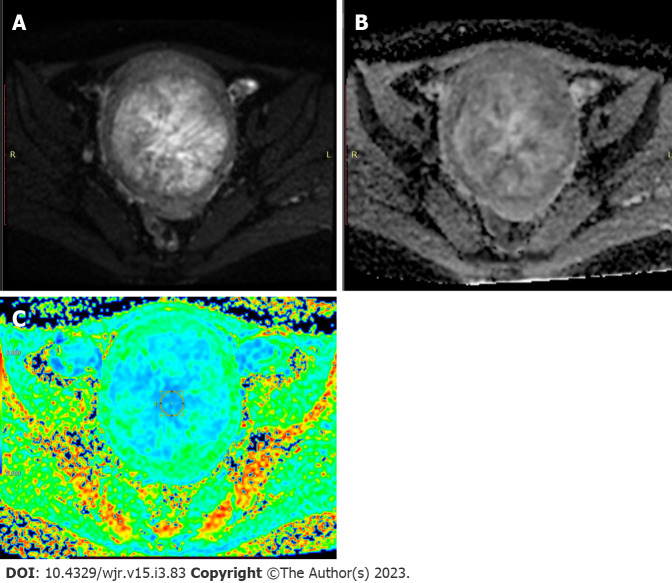
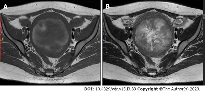
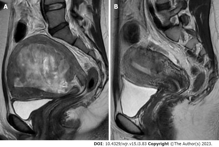
Case summary: Herein, we report a case of a pyomyoma in a nulliparous woman previously diagnosed with uterine leiomyomatosis according to ultrasound findings. The patient had previously attended the emergency room due to hypogastric pain unresponsive to analgesics. After a week of persistent pain, she developed sepsis without any identifiable foci. Magnetic resonance imaging revealed findings compatible with uterine myomatosis with red degeneration, and a possible diagnosis of a pyomyoma was made according to the imaging findings along with the patient's clinical features. We decided to perform myomectomy (which is an infrequently performed surgical treatment due to the procedure's intrinsic implications) due to the patient's desire to preserve fertility. Histopathologic results revealed a uterine leiomyoma with coagulative and liquefactive necrosis, while the tissue culture showed gram-negative cocci bacteria, which were successfully treated using antibiotic therapy. The patient's health status improved after several days.
Conclusion: The main diagnostic tools to evaluate pyomyomas are the clinical and laboratory findings as well as tissue cultures. Nonetheless, magnetic resonance imaging can help to corroborate these findings as well as to better characterize myomas with its different complications.


 分享
分享
 求助内容:
求助内容: 应助结果提醒方式:
 扫码关注我们
