{"title":"Fenestrated Anterior Communicating Artery Complex Mimicking an Unruptured Aneurysm: Diagnostic Pitfall.","authors":"Atsushi Tsukada, Kiyoyuki Yanaka, Hayato Takeda, Kuniyuki Onuma, Maya Takada, Kazuhiro Nakamura, Eiichi Ishikawa","doi":"10.1055/s-0043-1764119","DOIUrl":null,"url":null,"abstract":"<p><p>Anatomical variations often occur in the anterior communicating artery (AComA) complex, and a careful preoperative evaluation is required before repair of this lesion. We report a case of a fenestrated AComA complex mimicking an unruptured cerebral aneurysm. A 49-year-old woman was referred to our hospital under suspicion of unruptured aneurysms of the AComA and the left middle cerebral artery on magnetic resonance angiography (MRA). Additional three-dimensional computed tomographic angiography (CTA) showed the lesion arising from the AComA complex with a maximum diameter of 4.2 mm. Intraoperative findings showed that the putative aneurysm was actually a fenestrated AComA complex as the blood vessels that formed the AComA complex were dilated and meandering. After the operation, MRA and CTA three-dimensional images were reviewed again but we could still not diagnose the lesion as a fenestrated AComA complex rather than an aneurysm. However, in the MRA source image, a secant line in the lesion was the only finding suggestive of a fenestration. The AComA complex is often associated with various vascular malformations, and it is essential to consider this association in the preoperative evaluation. The interpretation of source images may be helpful for accurate diagnosis and surgical planning.</p>","PeriodicalId":8521,"journal":{"name":"Asian Journal of Neurosurgery","volume":"18 1","pages":"201-205"},"PeriodicalIF":0.0000,"publicationDate":"2023-03-01","publicationTypes":"Journal Article","fieldsOfStudy":null,"isOpenAccess":false,"openAccessPdf":"https://ftp.ncbi.nlm.nih.gov/pub/pmc/oa_pdf/88/0d/10-1055-s-0043-1764119.PMC10089750.pdf","citationCount":"0","resultStr":null,"platform":"Semanticscholar","paperid":null,"PeriodicalName":"Asian Journal of Neurosurgery","FirstCategoryId":"1085","ListUrlMain":"https://doi.org/10.1055/s-0043-1764119","RegionNum":0,"RegionCategory":null,"ArticlePicture":[],"TitleCN":null,"AbstractTextCN":null,"PMCID":null,"EPubDate":"","PubModel":"","JCR":"","JCRName":"","Score":null,"Total":0}
引用次数: 0
Abstract
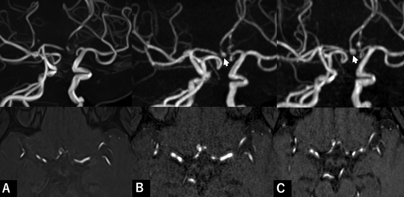
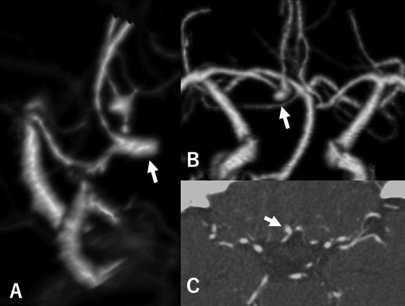
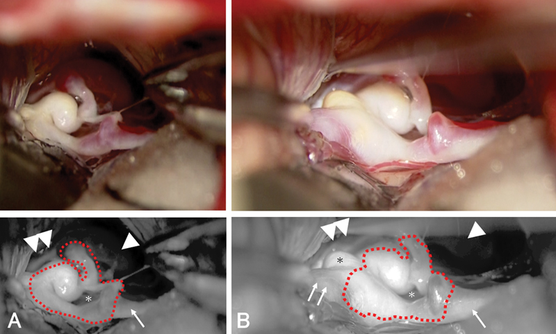
Anatomical variations often occur in the anterior communicating artery (AComA) complex, and a careful preoperative evaluation is required before repair of this lesion. We report a case of a fenestrated AComA complex mimicking an unruptured cerebral aneurysm. A 49-year-old woman was referred to our hospital under suspicion of unruptured aneurysms of the AComA and the left middle cerebral artery on magnetic resonance angiography (MRA). Additional three-dimensional computed tomographic angiography (CTA) showed the lesion arising from the AComA complex with a maximum diameter of 4.2 mm. Intraoperative findings showed that the putative aneurysm was actually a fenestrated AComA complex as the blood vessels that formed the AComA complex were dilated and meandering. After the operation, MRA and CTA three-dimensional images were reviewed again but we could still not diagnose the lesion as a fenestrated AComA complex rather than an aneurysm. However, in the MRA source image, a secant line in the lesion was the only finding suggestive of a fenestration. The AComA complex is often associated with various vascular malformations, and it is essential to consider this association in the preoperative evaluation. The interpretation of source images may be helpful for accurate diagnosis and surgical planning.


 分享
分享

 求助内容:
求助内容: 应助结果提醒方式:
 扫码关注我们
