D A Vorontsov, E V Gubarkova, M A Sirotkina, A A Sovetsky, A A Plekhanov, S S Kuznetsov, D A Davydova, A Yu Bogomolova, V Y Zaitsev, S V Gamayunov, A Y Vorontsov, V A Sobolevskiy, N D Gladkova
{"title":"Multimodal Optical Coherence Tomography for Intraoperative Evaluation of Tumor Margins and Surgical Margins in Breast-Conserving Surgery.","authors":"D A Vorontsov, E V Gubarkova, M A Sirotkina, A A Sovetsky, A A Plekhanov, S S Kuznetsov, D A Davydova, A Yu Bogomolova, V Y Zaitsev, S V Gamayunov, A Y Vorontsov, V A Sobolevskiy, N D Gladkova","doi":"10.17691/stm2022.14.2.03","DOIUrl":null,"url":null,"abstract":"<p><strong>The aim of the study: </strong>We compare the effectiveness of multimodal optical coherence tomography (MM OCT) in the traditional structural OCT mode and the OCT elastography (OCE) mode in addressing two clinically important tasks: (1) detecting groups of tumor cells at surgical margins during breast-сonserving surgery (BСS) in breast cancer (BC) and (2) identifying breast tumor margins. The obtained results were correlated with corresponding histological sections.</p><p><strong>Materials and methods: </strong>The study was performed on 100 surgical margin samples (top, bottom, medial, and lateral - four samples from each patient in total) obtained from 25 patients with BC who underwent BCS (lumpectomy), and on 25 postoperative tumor samples (to determine tumor margins). With MM OCT method, we visually and numerically assessed the scattering (level and depth of OCT signal penetration) and elastic (stiffness values, or Young's modulus (kPa)) properties of the tumor and non-tumor breast tissue and the obtained values were compared with the results of postoperative histological examination.</p><p><strong>Results: </strong>In 4 surgical margin samples (out of 100), with the OCE method we identified groups of histologically confirmed tumor cells (\"positive\" resection margins) at the distance of about 5 mm from the visible tumor margin. The identified zones were larger than 0.5 mm with stiffness of more than 400 kPa in all these cases. However, the structural OCT could not identify these groups of tumors and they were not distinguishable from the surrounding fibrous tissue.In the areas of tumor into non-tumor tissue transition, structural OCT images detected tumor margins only if they were adjacent to adipose tissue and did not detect them if there were adjacent to non-tumor fibrous tissue. OCE images with high stiffness values (more than 400 kPa) and high contrast showed a clear tumor margin with both adipose and fibrous tissue.</p><p><strong>Conclusion: </strong>The study demonstarets the potential of MM OCT, particularly its OCE mode, as a real-time method for intraoperative tumor margin and surgical margin assessment in BCS. OCE images compared to structural OCT images visualize higher contrast between different types of breast tissue (adipose tissue, fibrous stroma, hyalinized stroma, tumor cell clusters), as well as more accurate identification of the tumor border and detection of small groups of tumor cells at surgical margins. An algorithm for intraoperative MM OCT examination of the state of the resection margin is proposed in accordance with standard clinical guidelines for achieving clean surgical margins in breast cancer patients.</p>","PeriodicalId":51886,"journal":{"name":"Sovremennye Tehnologii v Medicine","volume":"14 2","pages":"26-38"},"PeriodicalIF":0.9000,"publicationDate":"2022-01-01","publicationTypes":"Journal Article","fieldsOfStudy":null,"isOpenAccess":false,"openAccessPdf":"https://www.ncbi.nlm.nih.gov/pmc/articles/PMC10090927/pdf/","citationCount":"2","resultStr":null,"platform":"Semanticscholar","paperid":null,"PeriodicalName":"Sovremennye Tehnologii v Medicine","FirstCategoryId":"1085","ListUrlMain":"https://doi.org/10.17691/stm2022.14.2.03","RegionNum":0,"RegionCategory":null,"ArticlePicture":[],"TitleCN":null,"AbstractTextCN":null,"PMCID":null,"EPubDate":"","PubModel":"","JCR":"Q4","JCRName":"MEDICINE, RESEARCH & EXPERIMENTAL","Score":null,"Total":0}
引用次数: 2
Abstract
The aim of the study: We compare the effectiveness of multimodal optical coherence tomography (MM OCT) in the traditional structural OCT mode and the OCT elastography (OCE) mode in addressing two clinically important tasks: (1) detecting groups of tumor cells at surgical margins during breast-сonserving surgery (BСS) in breast cancer (BC) and (2) identifying breast tumor margins. The obtained results were correlated with corresponding histological sections.
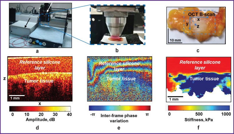
Materials and methods: The study was performed on 100 surgical margin samples (top, bottom, medial, and lateral - four samples from each patient in total) obtained from 25 patients with BC who underwent BCS (lumpectomy), and on 25 postoperative tumor samples (to determine tumor margins). With MM OCT method, we visually and numerically assessed the scattering (level and depth of OCT signal penetration) and elastic (stiffness values, or Young's modulus (kPa)) properties of the tumor and non-tumor breast tissue and the obtained values were compared with the results of postoperative histological examination.
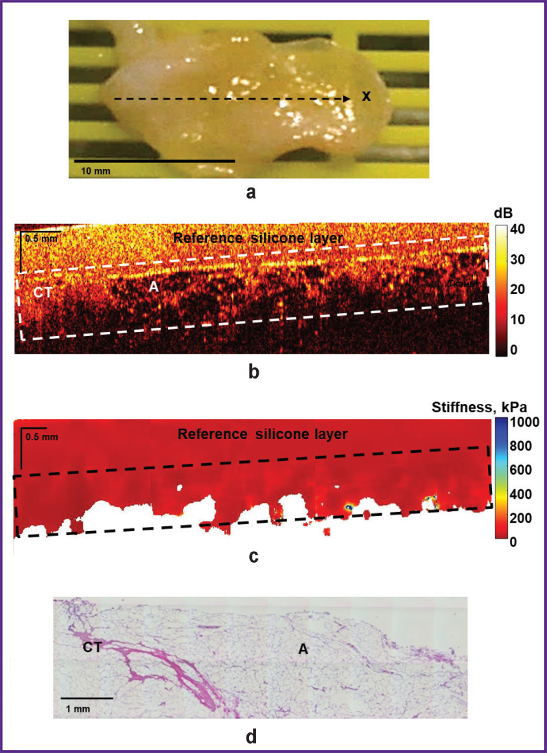
Results: In 4 surgical margin samples (out of 100), with the OCE method we identified groups of histologically confirmed tumor cells ("positive" resection margins) at the distance of about 5 mm from the visible tumor margin. The identified zones were larger than 0.5 mm with stiffness of more than 400 kPa in all these cases. However, the structural OCT could not identify these groups of tumors and they were not distinguishable from the surrounding fibrous tissue.In the areas of tumor into non-tumor tissue transition, structural OCT images detected tumor margins only if they were adjacent to adipose tissue and did not detect them if there were adjacent to non-tumor fibrous tissue. OCE images with high stiffness values (more than 400 kPa) and high contrast showed a clear tumor margin with both adipose and fibrous tissue.
Conclusion: The study demonstarets the potential of MM OCT, particularly its OCE mode, as a real-time method for intraoperative tumor margin and surgical margin assessment in BCS. OCE images compared to structural OCT images visualize higher contrast between different types of breast tissue (adipose tissue, fibrous stroma, hyalinized stroma, tumor cell clusters), as well as more accurate identification of the tumor border and detection of small groups of tumor cells at surgical margins. An algorithm for intraoperative MM OCT examination of the state of the resection margin is proposed in accordance with standard clinical guidelines for achieving clean surgical margins in breast cancer patients.


 分享
分享
 求助内容:
求助内容: 应助结果提醒方式:
 扫码关注我们
