{"title":"Repairing rat calvarial defects by adipose mesenchymal stem cells and novel freeze-dried three-dimensional nanofibrous scaffolds.","authors":"Maryam Sadat Khoramgah, Hossein Ghanbarian, Javad Ranjbari, Nilufar Ebrahimi, Fatemeh Sadat Tabatabaei Mirakabad, Navid Ahmady Roozbahany, Hojjat Allah Abbaszadeh, Simzar Hosseinzadeh","doi":"10.34172/bi.2021.23711","DOIUrl":null,"url":null,"abstract":"<p><p><b><i>Introduction:</i> </b> Treatment of critical-sized bone defects is challenging. Tissue engineering as a state-of-the-art method has been concerned with treating these non-self-healing bone defects. Here, we studied the potentials of new three-dimensional nanofibrous scaffolds (3DNS) with and without human adipose mesenchymal stem cells (ADSCs) for reconstructing rat critical-sized calvarial defects (CSCD). <i><b>Methods:</b> </i> Scaffolds were made from 1- polytetrafluoroethylene (PTFE), and polyvinyl alcohol (PVA) (PTFE/ PVA group), and 2- PTFE, PVA, and graphene oxide (GO) nanoparticle (PTFE/ PVA/GO group) and seeded by ADSCs and incubated in osteogenic media (OM). The expression of key osteogenic proteins including Runt-related transcription factor 2 (Runx2), collagen type Iα (COL Iα), osteocalcin (OCN), and osteonectin (ON) at days 14 and 21 of culture were evaluated by western blot and immunocytochemistry methods. Next, 40 selected rats were assigned to five groups (n=8) to create CSCD which will be filled by scaffolds or cell-containing scaffolds. The groups were denominated as the following order: Control (empty defects), PTFE/PVA (PTFE/PVA scaffolds implant), PTFE/PVA/GO (PTFE/PVA/GO scaffolds implant), PTFE/PVA/Cell group (PTFE/PVA scaffolds containing ADSCs implant), and PTFE/PVA/GO/Cell group (PTFE/PVA/GO scaffolds containing ADSCs implant). Six and 12 weeks after implantation, the animals were sacrificed and bone regeneration was evaluated using computerized tomography (CT), and hematoxylin-eosin (H&E) staining. <i><b>Results:</b> </i> Based on the in-vitro study, expression of bone-related proteins in ADSCs seeded on PTFE/PVA/GO scaffolds were significantly higher than PTFE/PVA scaffolds and TCPS (<i>P</i><0.05). Based on the in-vivo study, bone regeneration in CSCD were filled with PTFE/PVA/GO scaffolds containing ADSCs were significantly higher than PTFE/PVA scaffolds containing ADSCs (<i>P</i><0.05). CSCD filled with cell-seeded scaffolds showed higher bone regeneration in comparison with CSCD filled with scaffolds only (<i>P</i><0.05). <i><b>Conclusion:</b> </i> The data provided evidence showing new freeze-dried nanofibrous scaffolds formed from hydrophobic (PTFE) and hydrophilic (PVA) polymers with and without GO provide a suitable environment for ADSCs due to the expression of bone-related proteins. ADSCs and GO in the implanted scaffolds had a distinct effect on the bone regeneration process in this in-vivo study.</p>","PeriodicalId":48614,"journal":{"name":"Bioimpacts","volume":"13 1","pages":"31-42"},"PeriodicalIF":2.2000,"publicationDate":"2023-01-01","publicationTypes":"Journal Article","fieldsOfStudy":null,"isOpenAccess":false,"openAccessPdf":"https://ftp.ncbi.nlm.nih.gov/pub/pmc/oa_pdf/52/1c/bi-13-31.PMC9923815.pdf","citationCount":"2","resultStr":null,"platform":"Semanticscholar","paperid":null,"PeriodicalName":"Bioimpacts","FirstCategoryId":"5","ListUrlMain":"https://doi.org/10.34172/bi.2021.23711","RegionNum":4,"RegionCategory":"工程技术","ArticlePicture":[],"TitleCN":null,"AbstractTextCN":null,"PMCID":null,"EPubDate":"","PubModel":"","JCR":"Q3","JCRName":"PHARMACOLOGY & PHARMACY","Score":null,"Total":0}
引用次数: 2
Abstract
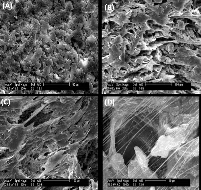
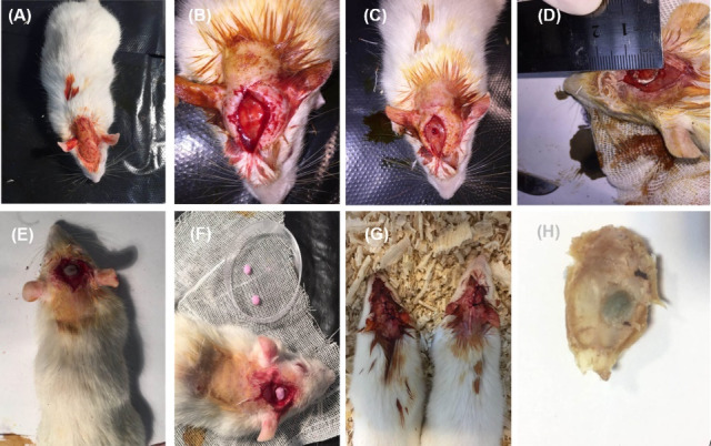
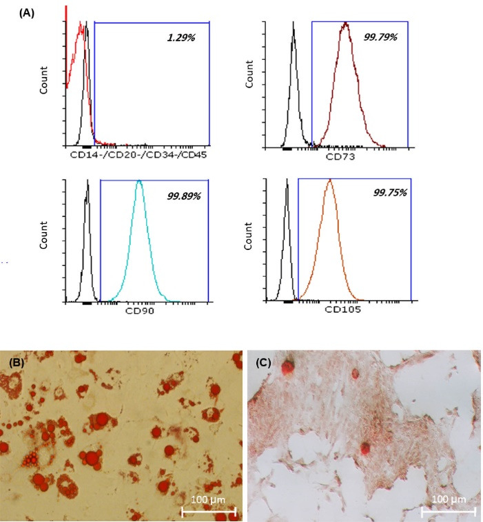
Introduction: Treatment of critical-sized bone defects is challenging. Tissue engineering as a state-of-the-art method has been concerned with treating these non-self-healing bone defects. Here, we studied the potentials of new three-dimensional nanofibrous scaffolds (3DNS) with and without human adipose mesenchymal stem cells (ADSCs) for reconstructing rat critical-sized calvarial defects (CSCD). Methods: Scaffolds were made from 1- polytetrafluoroethylene (PTFE), and polyvinyl alcohol (PVA) (PTFE/ PVA group), and 2- PTFE, PVA, and graphene oxide (GO) nanoparticle (PTFE/ PVA/GO group) and seeded by ADSCs and incubated in osteogenic media (OM). The expression of key osteogenic proteins including Runt-related transcription factor 2 (Runx2), collagen type Iα (COL Iα), osteocalcin (OCN), and osteonectin (ON) at days 14 and 21 of culture were evaluated by western blot and immunocytochemistry methods. Next, 40 selected rats were assigned to five groups (n=8) to create CSCD which will be filled by scaffolds or cell-containing scaffolds. The groups were denominated as the following order: Control (empty defects), PTFE/PVA (PTFE/PVA scaffolds implant), PTFE/PVA/GO (PTFE/PVA/GO scaffolds implant), PTFE/PVA/Cell group (PTFE/PVA scaffolds containing ADSCs implant), and PTFE/PVA/GO/Cell group (PTFE/PVA/GO scaffolds containing ADSCs implant). Six and 12 weeks after implantation, the animals were sacrificed and bone regeneration was evaluated using computerized tomography (CT), and hematoxylin-eosin (H&E) staining. Results: Based on the in-vitro study, expression of bone-related proteins in ADSCs seeded on PTFE/PVA/GO scaffolds were significantly higher than PTFE/PVA scaffolds and TCPS (P<0.05). Based on the in-vivo study, bone regeneration in CSCD were filled with PTFE/PVA/GO scaffolds containing ADSCs were significantly higher than PTFE/PVA scaffolds containing ADSCs (P<0.05). CSCD filled with cell-seeded scaffolds showed higher bone regeneration in comparison with CSCD filled with scaffolds only (P<0.05). Conclusion: The data provided evidence showing new freeze-dried nanofibrous scaffolds formed from hydrophobic (PTFE) and hydrophilic (PVA) polymers with and without GO provide a suitable environment for ADSCs due to the expression of bone-related proteins. ADSCs and GO in the implanted scaffolds had a distinct effect on the bone regeneration process in this in-vivo study.
BioimpactsPharmacology, Toxicology and Pharmaceutics-Pharmaceutical Science
CiteScore
4.80
自引率
7.70%
发文量
36
审稿时长
5 weeks
期刊介绍:
BioImpacts (BI) is a peer-reviewed multidisciplinary international journal, covering original research articles, reviews, commentaries, hypotheses, methodologies, and visions/reflections dealing with all aspects of biological and biomedical researches at molecular, cellular, functional and translational dimensions.


 分享
分享
 求助内容:
求助内容: 应助结果提醒方式:
 扫码关注我们
