Novel segmentation algorithm for high-throughput analysis of spectral domain-optical coherence tomography imaging of teleost retinas.
IF 1.4 3区 医学Q4 BIOCHEMISTRY & MOLECULAR BIOLOGYMolecular VisionPub Date : 2022-01-01
Kent R Barter, Hélène Paradis, Robert L Gendron, Josué A Lily Vidal, Oscar Meruvia-Pastor
{"title":"Novel segmentation algorithm for high-throughput analysis of spectral domain-optical coherence tomography imaging of teleost retinas.","authors":"Kent R Barter, Hélène Paradis, Robert L Gendron, Josué A Lily Vidal, Oscar Meruvia-Pastor","doi":"","DOIUrl":null,"url":null,"abstract":"<p><p>Spectral domain-optical coherence tomography (SD-OCT) has become an essential tool for assessing ocular tissues in live subjects and conducting research on ocular development, health, and disease. The processing of SD-OCT images, particularly those from non-mammalian species, is a labor-intensive manual process due to a lack of automated analytical programs. This paper describes the development and implementation of a novel computer algorithm for the quantitative analysis of SD-OCT images of live teleost eyes. Automated segmentation processing of SD-OCT images of retinal layers was developed using a novel algorithm based on thresholding. The algorithm measures retinal thickness characteristics in a large volume of imaging data of teleost ocular structures in a short time, providing increased accuracy and repeatability of SD-OCT image analysis over manual measurements. The algorithm also generates hundreds of retinal thickness measurements per image for a large number of images for a given dataset. Meanwhile, heat mapping software that plots SD-OCT image measurements as a color gradient was also created. This software directly converts the measurements of each processed image to represent changes in thickness across the whole retinal scan. It also enables 2D and 3D visualization of retinal thickness across the scan, facilitating specimen comparison and localization of areas of interest. The study findings showed that the novel algorithm is more accurate, reliable, and repeatable than manual SD-OCT analysis. The adaptability of the algorithm makes it potentially suitable for analyzing SD-OCT scans of other non-mammalian species.</p>","PeriodicalId":18866,"journal":{"name":"Molecular Vision","volume":"28 ","pages":"492-499"},"PeriodicalIF":1.4000,"publicationDate":"2022-01-01","publicationTypes":"Journal Article","fieldsOfStudy":null,"isOpenAccess":false,"openAccessPdf":"https://ftp.ncbi.nlm.nih.gov/pub/pmc/oa_pdf/4c/e6/mv-v28-492.PMC10115363.pdf","citationCount":"0","resultStr":null,"platform":"Semanticscholar","paperid":null,"PeriodicalName":"Molecular Vision","FirstCategoryId":"3","ListUrlMain":"","RegionNum":3,"RegionCategory":"医学","ArticlePicture":[],"TitleCN":null,"AbstractTextCN":null,"PMCID":null,"EPubDate":"","PubModel":"","JCR":"Q4","JCRName":"BIOCHEMISTRY & MOLECULAR BIOLOGY","Score":null,"Total":0}
引用次数: 0
Abstract
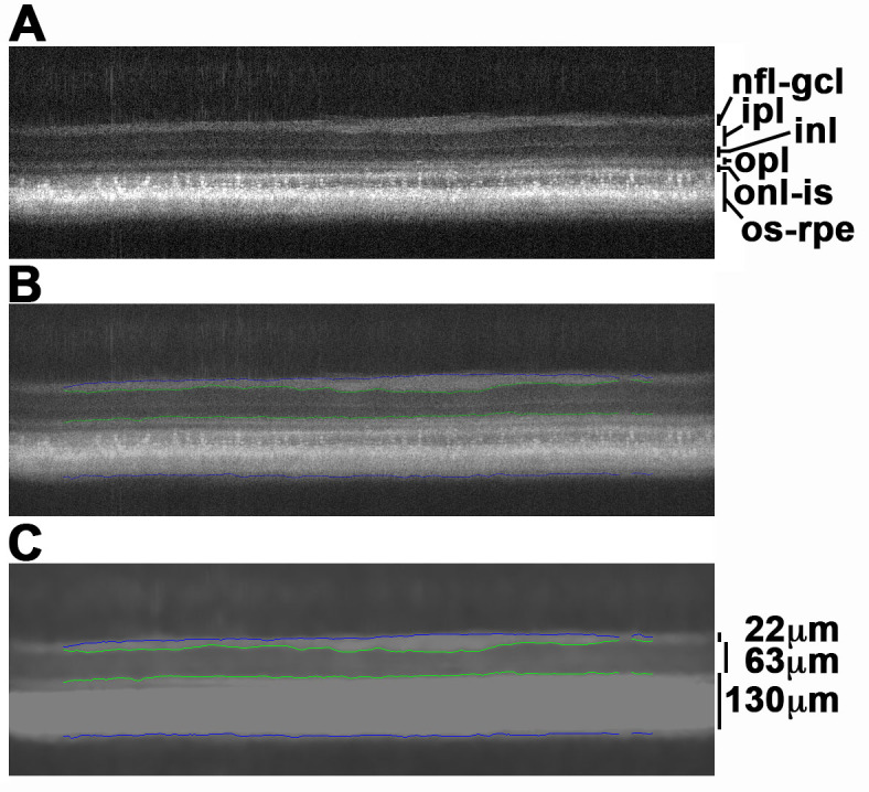
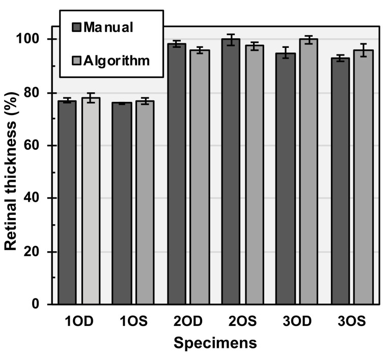
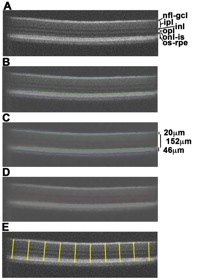
Spectral domain-optical coherence tomography (SD-OCT) has become an essential tool for assessing ocular tissues in live subjects and conducting research on ocular development, health, and disease. The processing of SD-OCT images, particularly those from non-mammalian species, is a labor-intensive manual process due to a lack of automated analytical programs. This paper describes the development and implementation of a novel computer algorithm for the quantitative analysis of SD-OCT images of live teleost eyes. Automated segmentation processing of SD-OCT images of retinal layers was developed using a novel algorithm based on thresholding. The algorithm measures retinal thickness characteristics in a large volume of imaging data of teleost ocular structures in a short time, providing increased accuracy and repeatability of SD-OCT image analysis over manual measurements. The algorithm also generates hundreds of retinal thickness measurements per image for a large number of images for a given dataset. Meanwhile, heat mapping software that plots SD-OCT image measurements as a color gradient was also created. This software directly converts the measurements of each processed image to represent changes in thickness across the whole retinal scan. It also enables 2D and 3D visualization of retinal thickness across the scan, facilitating specimen comparison and localization of areas of interest. The study findings showed that the novel algorithm is more accurate, reliable, and repeatable than manual SD-OCT analysis. The adaptability of the algorithm makes it potentially suitable for analyzing SD-OCT scans of other non-mammalian species.
期刊介绍:
Molecular Vision is a peer-reviewed journal dedicated to the dissemination of research results in molecular biology, cell biology, and the genetics of the visual system (ocular and cortical).
Molecular Vision publishes articles presenting original research that has not previously been published and comprehensive articles reviewing the current status of a particular field or topic. Submissions to Molecular Vision are subjected to rigorous peer review. Molecular Vision does NOT publish preprints.
For authors, Molecular Vision provides a rapid means of communicating important results. Access to Molecular Vision is free and unrestricted, allowing the widest possible audience for your article. Digital publishing allows you to use color images freely (and without fees). Additionally, you may publish animations, sounds, or other supplementary information that clarifies or supports your article. Each of the authors of an article may also list an electronic mail address (which will be updated upon request) to give interested readers easy access to authors.
 分享
分享


 求助内容:
求助内容: 应助结果提醒方式:
 扫码关注我们
