{"title":"Corneal perforation after corneal foreign body - Case Report.","authors":"Sorin Simion Macarie, Fodor Vlad, Daniela Mariana Macarie","doi":"10.22336/rjo.2023.14","DOIUrl":null,"url":null,"abstract":"<p><p><b>Purpose:</b> To present the case of a patient with a history of trauma and corneal foreign body in the right eye, followed by decreased visual acuity in the right eye, corneal perforation with good recovery after surgical treatment. <b>Material and method:</b> We report a case of a patient who presented to our clinic with a sudden decrease of visual acuity in the right eye, two months after an incident resulting in a corneal foreign body in the right eye. In the case presented, the patient applied a local medical self-treatment, an antibiotic and a topical corticosteroid. After a few weeks, the patient presented to the ophthalmologist, a foreign body was extracted from the cornea of the right eye and a topical treatment with a non-steroidal anti-inflammatory drug, a cycloplegic and an antibiotic were indicated. However, corneal perforation occurred and the patient was urgently sent to our service, where a corneal anaesthesia was also found. <b>Results:</b> Corneal perforation healed with a minor paracentral opacification. <b>Discussions:</b> Corneal perforation in our patient was due to corneal melting because of topical steroid anti-inflammatory autotherapy, late corneal foreign body extraction and topical treatment with non-steroidal anti-inflammatory drugs. Corneal anesthesia is also an important factor that enhances corneal melting and perforation. The surgical intervention performed healed the corneal perforation. <b>Conclusions:</b> Corneal anaesthesia and topical anti-inflammatory administration led to corneal perforation. Corneal sensitivity should be tested in patients with corneal foreign body. Corneal patching proved to be an adequate solution in this patient.</p>","PeriodicalId":21385,"journal":{"name":"Romanian journal of ophthalmology","volume":"67 1","pages":"77-80"},"PeriodicalIF":0.0000,"publicationDate":"2023-01-01","publicationTypes":"Journal Article","fieldsOfStudy":null,"isOpenAccess":false,"openAccessPdf":"https://www.ncbi.nlm.nih.gov/pmc/articles/PMC10117182/pdf/","citationCount":"0","resultStr":null,"platform":"Semanticscholar","paperid":null,"PeriodicalName":"Romanian journal of ophthalmology","FirstCategoryId":"1085","ListUrlMain":"https://doi.org/10.22336/rjo.2023.14","RegionNum":0,"RegionCategory":null,"ArticlePicture":[],"TitleCN":null,"AbstractTextCN":null,"PMCID":null,"EPubDate":"","PubModel":"","JCR":"","JCRName":"","Score":null,"Total":0}
引用次数: 0
Abstract
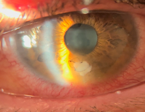
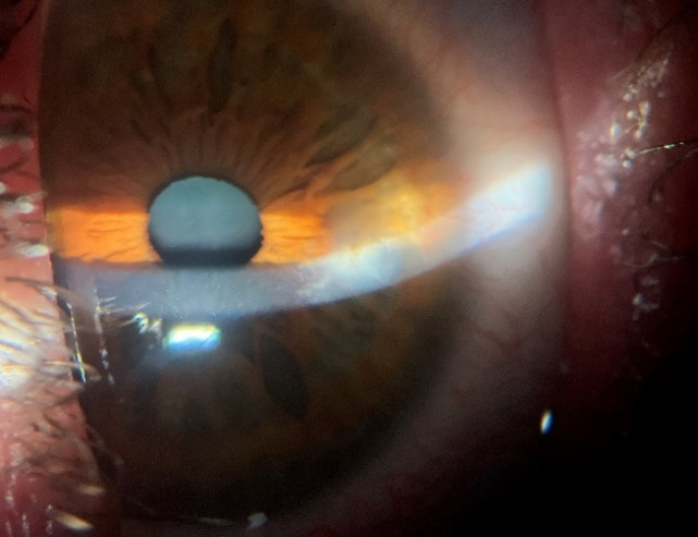
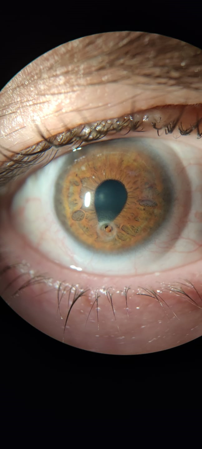
Purpose: To present the case of a patient with a history of trauma and corneal foreign body in the right eye, followed by decreased visual acuity in the right eye, corneal perforation with good recovery after surgical treatment. Material and method: We report a case of a patient who presented to our clinic with a sudden decrease of visual acuity in the right eye, two months after an incident resulting in a corneal foreign body in the right eye. In the case presented, the patient applied a local medical self-treatment, an antibiotic and a topical corticosteroid. After a few weeks, the patient presented to the ophthalmologist, a foreign body was extracted from the cornea of the right eye and a topical treatment with a non-steroidal anti-inflammatory drug, a cycloplegic and an antibiotic were indicated. However, corneal perforation occurred and the patient was urgently sent to our service, where a corneal anaesthesia was also found. Results: Corneal perforation healed with a minor paracentral opacification. Discussions: Corneal perforation in our patient was due to corneal melting because of topical steroid anti-inflammatory autotherapy, late corneal foreign body extraction and topical treatment with non-steroidal anti-inflammatory drugs. Corneal anesthesia is also an important factor that enhances corneal melting and perforation. The surgical intervention performed healed the corneal perforation. Conclusions: Corneal anaesthesia and topical anti-inflammatory administration led to corneal perforation. Corneal sensitivity should be tested in patients with corneal foreign body. Corneal patching proved to be an adequate solution in this patient.


 分享
分享

 求助内容:
求助内容: 应助结果提醒方式:
 扫码关注我们
