{"title":"Intravascular ultrasound imaging in evaluation of aortic stiffness: A proof-of-concept study.","authors":"Niya Boykova Mileva, Dobrin Iotkov Vassilev","doi":"10.5603/CJ.a2021.0003","DOIUrl":null,"url":null,"abstract":"<p><strong>Background: </strong>Aortic stiffness is a well-known cardio-vascular risk factor. For years, different methods have been studied in the assessment of aortic elastic properties and large arterial stiffness for risk stratification. Herein is an assessment of the role of intravascular ultrasound (IVUS) imaging for the evaluation of aortic elastic properties.</p><p><strong>Methods: </strong>Intravascular ultrasound imaging of the aorta was performed in 12 patients with transthoracic echocardiography (TTE) and computed tomography (CT) evidence for enlargement of the ascending aorta - diameter ≥ 40.0 mm. Mechanical properties of the aorta were derived from the measured diameters and intra-aortic pressure. Paired samples T-test analyses were performed to determine differences between measurements derived by TTE, CT and IVUS.</p><p><strong>Results: </strong>Mean values of the calculated elastic properties via IVUS of the ascending aorta were as follows: compliance 0.021 ± 0.02; strain 205 ± 4.3; aortic stiffness index 4.3 ± 0.75; elastic modulus 0.31 ± 0.05. On paired T-test analysis maximum ascending aortic diameter measured by CT aortography and IVUS did not differ significantly (t = -0.19, p = 0.985), but a significant difference between IVUS measurements and TTE derived diameters was found (t = 13.118, p = 0.034). On average, IVUS diameters were 2.3 mm larger than the results acquired by TTE (95% confidence interval: 14.21-17.13).</p><p><strong>Conclusions: </strong>Intravascular ultrasound examination of the ascending aorta provided larger diameters than the ones collected by means of TTE. However, IVUS measurements did not differ significantly from diameters derived by CT aortography.</p>","PeriodicalId":9492,"journal":{"name":"Cardiology journal","volume":"30 2","pages":"221-227"},"PeriodicalIF":1.9000,"publicationDate":"2023-01-01","publicationTypes":"Journal Article","fieldsOfStudy":null,"isOpenAccess":false,"openAccessPdf":"https://ftp.ncbi.nlm.nih.gov/pub/pmc/oa_pdf/5a/da/cardj-30-2-221.PMC10129260.pdf","citationCount":"0","resultStr":null,"platform":"Semanticscholar","paperid":null,"PeriodicalName":"Cardiology journal","FirstCategoryId":"3","ListUrlMain":"https://doi.org/10.5603/CJ.a2021.0003","RegionNum":3,"RegionCategory":"医学","ArticlePicture":[],"TitleCN":null,"AbstractTextCN":null,"PMCID":null,"EPubDate":"","PubModel":"","JCR":"Q2","JCRName":"CARDIAC & CARDIOVASCULAR SYSTEMS","Score":null,"Total":0}
引用次数: 0
Abstract
Background: Aortic stiffness is a well-known cardio-vascular risk factor. For years, different methods have been studied in the assessment of aortic elastic properties and large arterial stiffness for risk stratification. Herein is an assessment of the role of intravascular ultrasound (IVUS) imaging for the evaluation of aortic elastic properties.
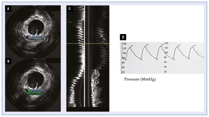
Methods: Intravascular ultrasound imaging of the aorta was performed in 12 patients with transthoracic echocardiography (TTE) and computed tomography (CT) evidence for enlargement of the ascending aorta - diameter ≥ 40.0 mm. Mechanical properties of the aorta were derived from the measured diameters and intra-aortic pressure. Paired samples T-test analyses were performed to determine differences between measurements derived by TTE, CT and IVUS.
Results: Mean values of the calculated elastic properties via IVUS of the ascending aorta were as follows: compliance 0.021 ± 0.02; strain 205 ± 4.3; aortic stiffness index 4.3 ± 0.75; elastic modulus 0.31 ± 0.05. On paired T-test analysis maximum ascending aortic diameter measured by CT aortography and IVUS did not differ significantly (t = -0.19, p = 0.985), but a significant difference between IVUS measurements and TTE derived diameters was found (t = 13.118, p = 0.034). On average, IVUS diameters were 2.3 mm larger than the results acquired by TTE (95% confidence interval: 14.21-17.13).
Conclusions: Intravascular ultrasound examination of the ascending aorta provided larger diameters than the ones collected by means of TTE. However, IVUS measurements did not differ significantly from diameters derived by CT aortography.
期刊介绍:
Cardiology Journal is a scientific, peer-reviewed journal covering a broad spectrum of topics in cardiology. The journal has been published since 1994 and over the years it has become an internationally recognized journal of cardiological and medical community.
Cardiology Journal is the journal for practicing cardiologists, researchers, and young trainees benefiting from broad spectrum of useful educational content.


 分享
分享
 求助内容:
求助内容: 应助结果提醒方式:
 扫码关注我们
