José C Candia, Osmar A Centurión, José F Alderete, Judith M Torales, Nelson J Aquino, Luis M Miño, Karina E Scavenius, Laura B García, Cristina Cáceres, Orlando J Sequeira, Christian O Chávez, Jorge E Martínez, Oscar A Lovera, E Javier Galeano
{"title":"Relationship of the T-wave Tpeak-Tend interval with conduction system disorders in arterial hypertension.","authors":"José C Candia, Osmar A Centurión, José F Alderete, Judith M Torales, Nelson J Aquino, Luis M Miño, Karina E Scavenius, Laura B García, Cristina Cáceres, Orlando J Sequeira, Christian O Chávez, Jorge E Martínez, Oscar A Lovera, E Javier Galeano","doi":"10.24875/ACM.21000396","DOIUrl":null,"url":null,"abstract":"<p><strong>Purpose: </strong>The Tpeak-Tend interval of the T wave has emerged as a new electrocardiographic marker of increased transmural dispersion of ventricular repolarization. We aimed to determine the presence of cardiac conduction system disorders in patients with systemic arterial hypertension (SAH) who have altered Tpeak-Tend interval of the T wave.</p><p><strong>Methods: </strong>The 67 patients with SAH were divided into two groups. Those with prolonged (≥ 77 ms) Tpeak-Tend intervals, 21 (31%) patients were in the study group. Those with normal (< 77 ms) Tpeak-Tend intervals, 46 (69%) patients were in the control group. Alteration of ventricular repolarization manifested as a prolongation of the Tpeak-Tend interval was detected by computerized electrocardiographic analysis tools.</p><p><strong>Results: </strong>The median value of QRS complex duration was significantly wider in the study group as compared to the control group (110 ± 12 ms vs. 94 ± 8 ms p < 0.001). There was a significantly greater incidence of left anterior hemiblock in the study group (14% vs. 0% p < 0.04). The median value of the QTc interval was significantly greater in the study group (440 ± 26 vs. 422 ± 15 p < 0.01). There was a significantly greater incidence of patients with prolonged QTc interval in the study group (33% vs. 11% p < 0.02). The median value of the Tpeak-Tend interval was significantly greater in the study group (84 ± 5 ms vs. 65 ± 4 ms p < 0.001), as well as, the Tpeak-Tend/QTc ratio in the study group (0.19 ± 0.1 vs. 0.16 ± 0.1 p < 0.001).</p><p><strong>Conclusion: </strong>There is a significantly greater ventricular repolarization disorders and abnormalities of the cardiac conduction system in SAH patients who possess altered Tpeak-Tend interval of the T wave.</p>","PeriodicalId":8360,"journal":{"name":"Archivos de cardiologia de Mexico","volume":"93 1","pages":"69-76"},"PeriodicalIF":0.7000,"publicationDate":"2023-01-01","publicationTypes":"Journal Article","fieldsOfStudy":null,"isOpenAccess":false,"openAccessPdf":"https://ftp.ncbi.nlm.nih.gov/pub/pmc/oa_pdf/c7/5c/7567AX221-ACM-93-69.PMC10161830.pdf","citationCount":"0","resultStr":null,"platform":"Semanticscholar","paperid":null,"PeriodicalName":"Archivos de cardiologia de Mexico","FirstCategoryId":"1085","ListUrlMain":"https://doi.org/10.24875/ACM.21000396","RegionNum":0,"RegionCategory":null,"ArticlePicture":[],"TitleCN":null,"AbstractTextCN":null,"PMCID":null,"EPubDate":"","PubModel":"","JCR":"Q4","JCRName":"CARDIAC & CARDIOVASCULAR SYSTEMS","Score":null,"Total":0}
引用次数: 0
Abstract
Purpose: The Tpeak-Tend interval of the T wave has emerged as a new electrocardiographic marker of increased transmural dispersion of ventricular repolarization. We aimed to determine the presence of cardiac conduction system disorders in patients with systemic arterial hypertension (SAH) who have altered Tpeak-Tend interval of the T wave.
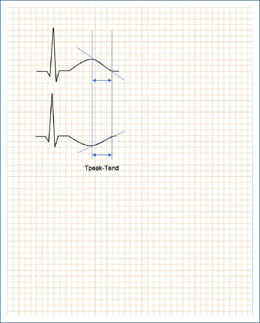
Methods: The 67 patients with SAH were divided into two groups. Those with prolonged (≥ 77 ms) Tpeak-Tend intervals, 21 (31%) patients were in the study group. Those with normal (< 77 ms) Tpeak-Tend intervals, 46 (69%) patients were in the control group. Alteration of ventricular repolarization manifested as a prolongation of the Tpeak-Tend interval was detected by computerized electrocardiographic analysis tools.
Results: The median value of QRS complex duration was significantly wider in the study group as compared to the control group (110 ± 12 ms vs. 94 ± 8 ms p < 0.001). There was a significantly greater incidence of left anterior hemiblock in the study group (14% vs. 0% p < 0.04). The median value of the QTc interval was significantly greater in the study group (440 ± 26 vs. 422 ± 15 p < 0.01). There was a significantly greater incidence of patients with prolonged QTc interval in the study group (33% vs. 11% p < 0.02). The median value of the Tpeak-Tend interval was significantly greater in the study group (84 ± 5 ms vs. 65 ± 4 ms p < 0.001), as well as, the Tpeak-Tend/QTc ratio in the study group (0.19 ± 0.1 vs. 0.16 ± 0.1 p < 0.001).
Conclusion: There is a significantly greater ventricular repolarization disorders and abnormalities of the cardiac conduction system in SAH patients who possess altered Tpeak-Tend interval of the T wave.
目的:T波的峰值-倾向间隔已成为心室复极跨壁弥散度增加的一种新的心电图标志。我们的目的是确定T波Tpeak-Tend间隔改变的全身性动脉高血压(SAH)患者是否存在心脏传导系统障碍。方法:将67例SAH患者分为两组。Tpeak-Tend时间间隔延长(≥77 ms)者,研究组21例(31%)。Tpeak-Tend间隔正常(< 77 ms)者,46例(69%)为对照组。通过计算机心电图分析工具检测到心室复极的改变,表现为Tpeak-Tend间期的延长。结果:与对照组相比,研究组QRS复合持续时间的中位数明显更宽(110±12 ms vs 94±8 ms p < 0.001)。研究组左前叶阻滞的发生率明显高于对照组(14% vs. 0% p < 0.04)。研究组QTc间隔的中位值显著高于对照组(440±26比422±15 p < 0.01)。研究组QTc间期延长的发生率显著高于对照组(33% vs. 11% p < 0.02)。研究组Tpeak-Tend间隔的中位数(84±5 ms vs. 65±4 ms p < 0.001)和Tpeak-Tend/QTc比值(0.19±0.1 vs. 0.16±0.1 p < 0.001)显著高于研究组。结论:伴有T波Tpeak-Tend间期改变的SAH患者存在更大的心室复极障碍和心传导系统异常。


 分享
分享
 求助内容:
求助内容: 应助结果提醒方式:
 扫码关注我们
