Jong Hyun Park, Dong Hee Kang, Hong Bae Jeon, Hyonsurk Kim
{"title":"Orbital wall restoration with primary bone fragments in complex orbital fractures.","authors":"Jong Hyun Park, Dong Hee Kang, Hong Bae Jeon, Hyonsurk Kim","doi":"10.7181/acfs.2022.01116","DOIUrl":null,"url":null,"abstract":"<p><strong>Background: </strong>Complex orbital fractures are impure orbital fractures because they involve the orbital walls and mid-facial bones. The author reported an orbital wall restoration technique in which the primary orbital wall fragments were restored to their prior position in complex orbital fractures in 2020. As a follow-up to a previous preliminary study, this study retrospectively reviewed the surgical results of complex orbital wall fractures over a 4-year period and compared the surgical outcomes by dividing them into groups with and without balloon restoration.</p><p><strong>Methods: </strong>Data of 939 patients with facial bone fractures between August 2018 and August 2022 were reviewed. Of these, 154 had complex orbital fractures. Among them, 44 and 110 underwent reduction with and without the balloon technique respectively. Pre- and postoperative Naugle exophthalmometer (Good-Lite Co.) scales were evaluated. The orbital volume and orbital volume ratio were calculated from preoperative and 6 months postoperative computed tomography images.</p><p><strong>Results: </strong>Among 154 patients with complex orbital fractures, 44 patients underwent restoration with the balloon technique, and 110 patients underwent restoration without it. The Naugle scale did not differ significantly between the two groups, but the orbital volume ratio significantly decreased by 3.32% and 2.39% in groups with and without the balloon technique and the difference in OVR was significantly greater in patients in the balloon restoration group compared with the control group. Postoperative balloon rupture occurred in six out of 44 cases (13.64%). None of the six patients with balloon rupture showed significant enophthalmos at 6 months of follow-up.</p><p><strong>Conclusion: </strong>The balloon rupture rate was 13.64% (6/44 cases) with marginal screw fixation, blunt screws, and extra protection with a resorbable foam dressing. Furthermore, we restored the orbital wall with primary orbital fragments using balloon support in complex orbital wall fractures.</p>","PeriodicalId":52238,"journal":{"name":"Archives of Craniofacial Surgery","volume":"24 2","pages":"52-58"},"PeriodicalIF":0.0000,"publicationDate":"2023-04-01","publicationTypes":"Journal Article","fieldsOfStudy":null,"isOpenAccess":false,"openAccessPdf":"https://ftp.ncbi.nlm.nih.gov/pub/pmc/oa_pdf/48/ed/acfs-2022-01116.PMC10165238.pdf","citationCount":"1","resultStr":null,"platform":"Semanticscholar","paperid":null,"PeriodicalName":"Archives of Craniofacial Surgery","FirstCategoryId":"1085","ListUrlMain":"https://doi.org/10.7181/acfs.2022.01116","RegionNum":0,"RegionCategory":null,"ArticlePicture":[],"TitleCN":null,"AbstractTextCN":null,"PMCID":null,"EPubDate":"","PubModel":"","JCR":"Q2","JCRName":"Medicine","Score":null,"Total":0}
引用次数: 1
Abstract
Background: Complex orbital fractures are impure orbital fractures because they involve the orbital walls and mid-facial bones. The author reported an orbital wall restoration technique in which the primary orbital wall fragments were restored to their prior position in complex orbital fractures in 2020. As a follow-up to a previous preliminary study, this study retrospectively reviewed the surgical results of complex orbital wall fractures over a 4-year period and compared the surgical outcomes by dividing them into groups with and without balloon restoration.
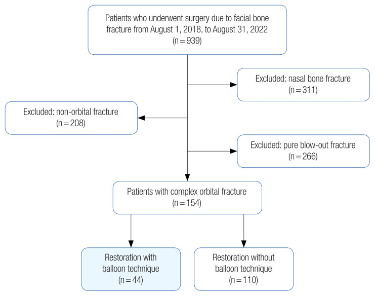
Methods: Data of 939 patients with facial bone fractures between August 2018 and August 2022 were reviewed. Of these, 154 had complex orbital fractures. Among them, 44 and 110 underwent reduction with and without the balloon technique respectively. Pre- and postoperative Naugle exophthalmometer (Good-Lite Co.) scales were evaluated. The orbital volume and orbital volume ratio were calculated from preoperative and 6 months postoperative computed tomography images.
Results: Among 154 patients with complex orbital fractures, 44 patients underwent restoration with the balloon technique, and 110 patients underwent restoration without it. The Naugle scale did not differ significantly between the two groups, but the orbital volume ratio significantly decreased by 3.32% and 2.39% in groups with and without the balloon technique and the difference in OVR was significantly greater in patients in the balloon restoration group compared with the control group. Postoperative balloon rupture occurred in six out of 44 cases (13.64%). None of the six patients with balloon rupture showed significant enophthalmos at 6 months of follow-up.
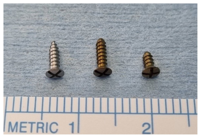
Conclusion: The balloon rupture rate was 13.64% (6/44 cases) with marginal screw fixation, blunt screws, and extra protection with a resorbable foam dressing. Furthermore, we restored the orbital wall with primary orbital fragments using balloon support in complex orbital wall fractures.


 分享
分享
 求助内容:
求助内容: 应助结果提醒方式:
 扫码关注我们
