{"title":"Endodontic Management of Maxillary Central Incisor with Two Roots, and Lateral Incisor with a C-shaped Canal; A Case Report.","authors":"Mageshwari Mahadevan, Benin Paulaian, Santhakumari Madhavankutty Ravisankar, Alexander Arvind Kumar, Neelamani Jaya Nagaraj","doi":"10.22037/iej.v18i2.38146","DOIUrl":null,"url":null,"abstract":"<p><p>The knowledge of anatomical variations in the morphology of root canal systems can affect the successful diagnosis to deliver proper endodontic treatment. The current case report enlightens the endodontic management of an anomalous maxillary left central incisor with two roots/root canals, a C-shaped root canal configuration in a maxillary left lateral incisor identified by three-dimensional cone-beam computed tomography imaging as well as the successful aesthetic rehabilitation of maxillary fractured incisors. The chief complaint of patient was a history of trauma during his outdoor play and consequent broken upper front teeth. Tooth #9 was diagnosed with pulpal necrosis accompanied by asymptomatic apical periodontitis with two relatively dilacerated roots while the maxillary left lateral incisor (tooth #10) was diagnosed with necrotic pulp and asymptomatic apical periodontitis having a C-shaped canal. Endodontic treatment for teeth #9 and #10 were performed, followed by post and core fabrication. Tooth reinforcement was achieved with prefabricated un-polymerized glass fiber post for lateral incisor and Interlig Fiber for central incisor. Intentional root canal treatment of tooth #8 was considered to reduce labial inclination. The anomalous maxillary central incisor with two roots is an unexpected variant during endodontic treatment, and the presence of C-shaped canal in lateral incisors is extremely rare requiring careful diagnosis with radiographs, clinical examination along with additional aids; <i>e.g.</i> Three-dimensional (3-D) cone-beam computed tomography. 3-D imaging has added the advantages of appropriate identification of anomalous anterior teeth and careful location of additional root canal(s) during endodontic treatment.</p>","PeriodicalId":14534,"journal":{"name":"Iranian Endodontic Journal","volume":"18 2","pages":"104-109"},"PeriodicalIF":0.0000,"publicationDate":"2023-01-01","publicationTypes":"Journal Article","fieldsOfStudy":null,"isOpenAccess":false,"openAccessPdf":"https://ftp.ncbi.nlm.nih.gov/pub/pmc/oa_pdf/f8/8d/IEJ-18-104.PMC10155102.pdf","citationCount":"2","resultStr":null,"platform":"Semanticscholar","paperid":null,"PeriodicalName":"Iranian Endodontic Journal","FirstCategoryId":"1085","ListUrlMain":"https://doi.org/10.22037/iej.v18i2.38146","RegionNum":0,"RegionCategory":null,"ArticlePicture":[],"TitleCN":null,"AbstractTextCN":null,"PMCID":null,"EPubDate":"","PubModel":"","JCR":"Q3","JCRName":"Dentistry","Score":null,"Total":0}
引用次数: 2
Abstract
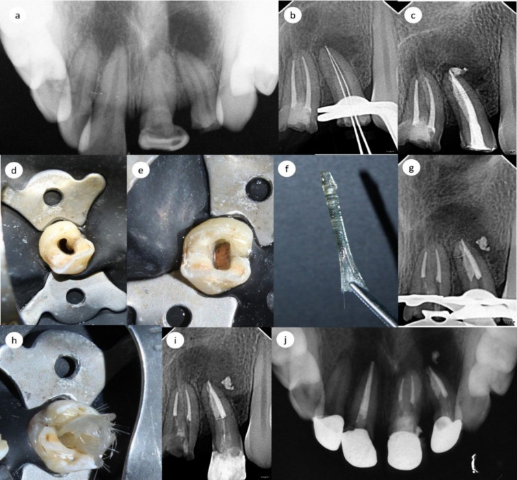
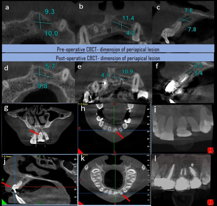
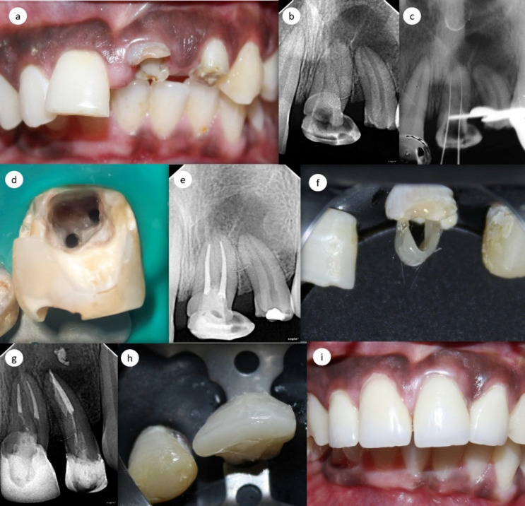
The knowledge of anatomical variations in the morphology of root canal systems can affect the successful diagnosis to deliver proper endodontic treatment. The current case report enlightens the endodontic management of an anomalous maxillary left central incisor with two roots/root canals, a C-shaped root canal configuration in a maxillary left lateral incisor identified by three-dimensional cone-beam computed tomography imaging as well as the successful aesthetic rehabilitation of maxillary fractured incisors. The chief complaint of patient was a history of trauma during his outdoor play and consequent broken upper front teeth. Tooth #9 was diagnosed with pulpal necrosis accompanied by asymptomatic apical periodontitis with two relatively dilacerated roots while the maxillary left lateral incisor (tooth #10) was diagnosed with necrotic pulp and asymptomatic apical periodontitis having a C-shaped canal. Endodontic treatment for teeth #9 and #10 were performed, followed by post and core fabrication. Tooth reinforcement was achieved with prefabricated un-polymerized glass fiber post for lateral incisor and Interlig Fiber for central incisor. Intentional root canal treatment of tooth #8 was considered to reduce labial inclination. The anomalous maxillary central incisor with two roots is an unexpected variant during endodontic treatment, and the presence of C-shaped canal in lateral incisors is extremely rare requiring careful diagnosis with radiographs, clinical examination along with additional aids; e.g. Three-dimensional (3-D) cone-beam computed tomography. 3-D imaging has added the advantages of appropriate identification of anomalous anterior teeth and careful location of additional root canal(s) during endodontic treatment.
期刊介绍:
The Iranian Endodontic Journal (IEJ) is an international peer-reviewed biomedical publication, the aim of which is to provide a scientific medium of communication for researchers throughout the globe. IEJ aims to publish the highest quality articles, both clinical and scientific, on all aspects of Endodontics. The journal is an official Journal of the Iranian Center for Endodontic Research (ICER) and the Iranian Association of Endodontists (IAE). The Journal welcomes articles related to the scientific or applied aspects of endodontics e.g. original researches, systematic reviews, meta-analyses, review articles, clinical trials, case series/reports, hypotheses, letters to the editor, etc. From the beginning (i.e. since 2006), the IEJ was the first open access endodontic journal in the world, which gave readers free and instant access to published articles and enabling them faster discovery of the latest endodontic research.


 分享
分享
 求助内容:
求助内容: 应助结果提醒方式:
 扫码关注我们
