Maria Rubega, Massimiliano Facca, Vittorio Curci, Giovanni Sparacino, Franco Molteni, Eleonora Guanziroli, Stefano Masiero, Emanuela Formaggio, Alessandra Del Felice
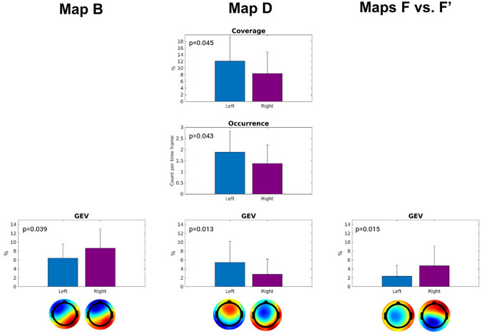
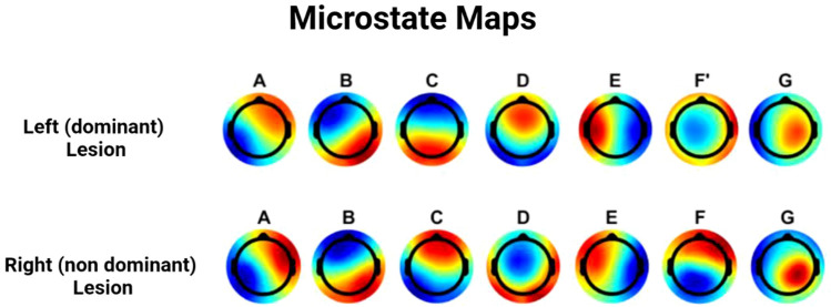
{"title":"EEG Microstates as a Signature of Hemispheric Lateralization in Stroke.","authors":"Maria Rubega, Massimiliano Facca, Vittorio Curci, Giovanni Sparacino, Franco Molteni, Eleonora Guanziroli, Stefano Masiero, Emanuela Formaggio, Alessandra Del Felice","doi":"10.1007/s10548-023-00967-8","DOIUrl":null,"url":null,"abstract":"<p><p>Stroke recovery trajectories vary substantially. The need for tracking and prognostic biomarkers in stroke is utmost for prognostic and rehabilitative goals: electroencephalography (EEG) advanced signal analysis may provide useful tools toward this aim. EEG microstates quantify changes in configuration of neuronal generators of short-lasting periods of coordinated synchronized communication within large-scale brain networks: this feature is expected to be impaired in stroke. To characterize the spatio-temporal signatures of EEG microstates in stroke survivors in the acute/subacute phase, EEG microstate analysis was performed in 51 first-ever ischemic stroke survivors [(28-82) years, 24 with right hemisphere (RH) lesion] who underwent a resting-state EEG recording in the acute and subacute phase (from 48 h up to 42 days after the event). Microstates were characterized based on 4 parameters: global explained variance (GEV), mean duration, occurrences per second, and percentage of coverage. Wilcoxon Rank Sum tests were performed to compare features of each microstate across the two groups [i.e., left hemisphere (LH) and right hemisphere (RH) stroke survivors]. The canonical microstate map D, characterized by a mostly frontal topography, displayed greater GEV, occurrence per second, and percentage of coverage in LH than in RH stroke survivors (p < 0.05). The EEG microstate map B, with a left-frontal to right-posterior topography, and F, with an occipital-to-frontal topography, exhibited a greater GEV in RH than in LH stroke survivors (p = 0.015). EEG microstates identified specific topographic maps which characterize stroke survivors' lesioned hemisphere in the acute and early subacute phase. Microstate features offer an additional tool to identify different neural reorganization.</p>","PeriodicalId":55329,"journal":{"name":"Brain Topography","volume":" ","pages":"475-478"},"PeriodicalIF":2.9000,"publicationDate":"2024-05-01","publicationTypes":"Journal Article","fieldsOfStudy":null,"isOpenAccess":false,"openAccessPdf":"https://www.ncbi.nlm.nih.gov/pmc/articles/PMC10191079/pdf/","citationCount":"0","resultStr":null,"platform":"Semanticscholar","paperid":null,"PeriodicalName":"Brain Topography","FirstCategoryId":"3","ListUrlMain":"https://doi.org/10.1007/s10548-023-00967-8","RegionNum":3,"RegionCategory":"医学","ArticlePicture":[],"TitleCN":null,"AbstractTextCN":null,"PMCID":null,"EPubDate":"2023/5/17 0:00:00","PubModel":"Epub","JCR":"Q3","JCRName":"CLINICAL NEUROLOGY","Score":null,"Total":0}
引用次数: 0
Abstract
Stroke recovery trajectories vary substantially. The need for tracking and prognostic biomarkers in stroke is utmost for prognostic and rehabilitative goals: electroencephalography (EEG) advanced signal analysis may provide useful tools toward this aim. EEG microstates quantify changes in configuration of neuronal generators of short-lasting periods of coordinated synchronized communication within large-scale brain networks: this feature is expected to be impaired in stroke. To characterize the spatio-temporal signatures of EEG microstates in stroke survivors in the acute/subacute phase, EEG microstate analysis was performed in 51 first-ever ischemic stroke survivors [(28-82) years, 24 with right hemisphere (RH) lesion] who underwent a resting-state EEG recording in the acute and subacute phase (from 48 h up to 42 days after the event). Microstates were characterized based on 4 parameters: global explained variance (GEV), mean duration, occurrences per second, and percentage of coverage. Wilcoxon Rank Sum tests were performed to compare features of each microstate across the two groups [i.e., left hemisphere (LH) and right hemisphere (RH) stroke survivors]. The canonical microstate map D, characterized by a mostly frontal topography, displayed greater GEV, occurrence per second, and percentage of coverage in LH than in RH stroke survivors (p < 0.05). The EEG microstate map B, with a left-frontal to right-posterior topography, and F, with an occipital-to-frontal topography, exhibited a greater GEV in RH than in LH stroke survivors (p = 0.015). EEG microstates identified specific topographic maps which characterize stroke survivors' lesioned hemisphere in the acute and early subacute phase. Microstate features offer an additional tool to identify different neural reorganization.
期刊介绍:
Brain Topography publishes clinical and basic research on cognitive neuroscience and functional neurophysiology using the full range of imaging techniques including EEG, MEG, fMRI, TMS, diffusion imaging, spectroscopy, intracranial recordings, lesion studies, and related methods. Submissions combining multiple techniques are particularly encouraged, as well as reports of new and innovative methodologies.


 分享
分享
 求助内容:
求助内容: 应助结果提醒方式:
 扫码关注我们
