Davaine Joel Ndongo Sonfack, Bilal Tarabay, Jesse Shen, Zhi Wang, Ghassan Boubez, Daniel Shédid, Sung-Joo Yuh
{"title":"Pneumorrhachis and pneumocephalus resulting from pneumothorax: illustrative case.","authors":"Davaine Joel Ndongo Sonfack, Bilal Tarabay, Jesse Shen, Zhi Wang, Ghassan Boubez, Daniel Shédid, Sung-Joo Yuh","doi":"10.3171/CASE23129","DOIUrl":null,"url":null,"abstract":"<p><strong>Background: </strong>Pneumorrhachis and pneumocephalus are rare conditions in which air is found within the spinal canal and brain, respectively. It is mostly asymptomatic and can be located in the intradural or extradural space. Intradural pneumorrhachis should prompt clinicians to search and treat any underlying injury of the skull, chest, or spinal column.</p><p><strong>Observations: </strong>A 68-year-old man presented with a history of cardiopulmonary arrest together with pneumorrhachis and pneumocephalus following a recurrent pneumothorax. The patient reported acute headaches with no other neurological symptoms. He was managed conservatively with bed rest for 48 hours following thoracoscopic talcage of his pneumothorax. Follow-up imaging showed regression of the pneumorrhachis, and the patient reported no other neurological symptoms.</p><p><strong>Lessons: </strong>Pneumorrhachis is an incidental radiological finding that self-resolves with conservative management. However, it can be a complication resulting from a serious injury. Therefore, close monitoring of neurological symptoms and complete investigations should be performed in patients with pneumorrhachis.</p>","PeriodicalId":16554,"journal":{"name":"Journal of Neurosurgery: Case Lessons","volume":"5 20","pages":""},"PeriodicalIF":0.0000,"publicationDate":"2023-05-15","publicationTypes":"Journal Article","fieldsOfStudy":null,"isOpenAccess":false,"openAccessPdf":"https://ftp.ncbi.nlm.nih.gov/pub/pmc/oa_pdf/e7/ce/CASE23129.PMC10550525.pdf","citationCount":"0","resultStr":null,"platform":"Semanticscholar","paperid":null,"PeriodicalName":"Journal of Neurosurgery: Case Lessons","FirstCategoryId":"1085","ListUrlMain":"https://doi.org/10.3171/CASE23129","RegionNum":0,"RegionCategory":null,"ArticlePicture":[],"TitleCN":null,"AbstractTextCN":null,"PMCID":null,"EPubDate":"","PubModel":"","JCR":"","JCRName":"","Score":null,"Total":0}
引用次数: 0
Abstract
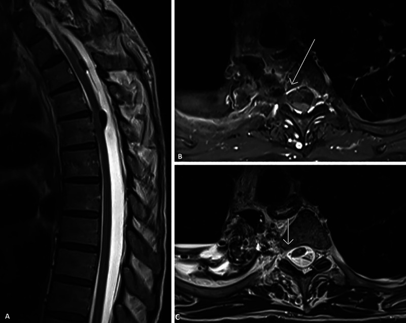
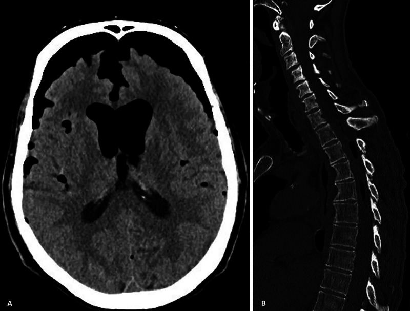
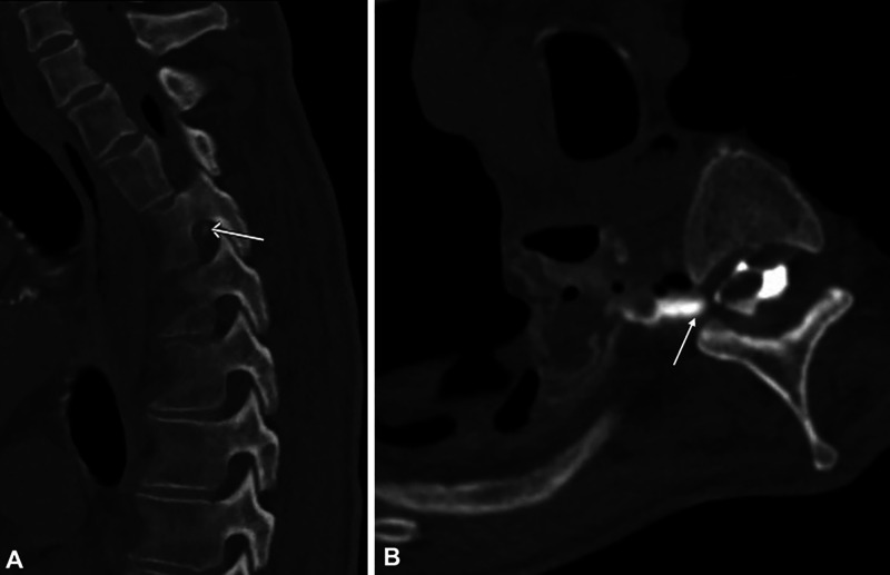
Background: Pneumorrhachis and pneumocephalus are rare conditions in which air is found within the spinal canal and brain, respectively. It is mostly asymptomatic and can be located in the intradural or extradural space. Intradural pneumorrhachis should prompt clinicians to search and treat any underlying injury of the skull, chest, or spinal column.
Observations: A 68-year-old man presented with a history of cardiopulmonary arrest together with pneumorrhachis and pneumocephalus following a recurrent pneumothorax. The patient reported acute headaches with no other neurological symptoms. He was managed conservatively with bed rest for 48 hours following thoracoscopic talcage of his pneumothorax. Follow-up imaging showed regression of the pneumorrhachis, and the patient reported no other neurological symptoms.
Lessons: Pneumorrhachis is an incidental radiological finding that self-resolves with conservative management. However, it can be a complication resulting from a serious injury. Therefore, close monitoring of neurological symptoms and complete investigations should be performed in patients with pneumorrhachis.


 分享
分享

 求助内容:
求助内容: 应助结果提醒方式:
 扫码关注我们
