Jin Woon Jeong, Ji Hyun Noh, Jeong Hyun Kang, Ji Hyun Park, Joo Hyung Lee
{"title":"Cecal malakoplakia: A case report.","authors":"Jin Woon Jeong, Ji Hyun Noh, Jeong Hyun Kang, Ji Hyun Park, Joo Hyung Lee","doi":"10.14216/kjco.21007","DOIUrl":null,"url":null,"abstract":"<p><p>Malakoplakia is a rare chronic granulomatous disease found in the genitourinary tract, mainly. It is considered to be related to immunosuppression and/or infectious processes. We would like to present an operative case of cecal malakoplakia in a patient with a history of surgical resection and chemotherapy for cervical cancer. A 74-year-old female patient visited our hospital for 1-year follow-up after operation and chemo-radiotherapy for cervical cancer. An infiltrative mass of 6 cm, between the cecal base and the right psoas muscle, was observed on computed tomography. An ileocectomy was performed for diagnosis. Histopathologic examination revealed cecal malakoplakia. After surgery, based on previous reports, antibiotics therapy was added. Then the patient was discharged and treated in the outpatient clinic. To our knowledge, a rare case has been described of cecal malakoplakia during observation after surgery and chemo-radiotherapy for cervical cancer. Malakoplakia is known to be related to immunosuppressive condition. Therefore, our case suggests that close observation should be made in patients on immunosuppressive condition, such as chemotherapy.</p>","PeriodicalId":74045,"journal":{"name":"Korean journal of clinical oncology","volume":"17 1","pages":"44-47"},"PeriodicalIF":0.0000,"publicationDate":"2021-06-01","publicationTypes":"Journal Article","fieldsOfStudy":null,"isOpenAccess":false,"openAccessPdf":"https://ftp.ncbi.nlm.nih.gov/pub/pmc/oa_pdf/18/d1/kjco-17-1-44.PMC9942739.pdf","citationCount":"0","resultStr":null,"platform":"Semanticscholar","paperid":null,"PeriodicalName":"Korean journal of clinical oncology","FirstCategoryId":"1085","ListUrlMain":"https://doi.org/10.14216/kjco.21007","RegionNum":0,"RegionCategory":null,"ArticlePicture":[],"TitleCN":null,"AbstractTextCN":null,"PMCID":null,"EPubDate":"","PubModel":"","JCR":"","JCRName":"","Score":null,"Total":0}
引用次数: 0
Abstract
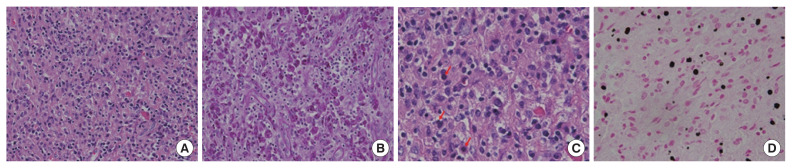
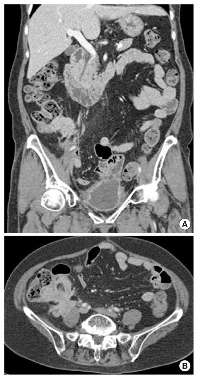
Malakoplakia is a rare chronic granulomatous disease found in the genitourinary tract, mainly. It is considered to be related to immunosuppression and/or infectious processes. We would like to present an operative case of cecal malakoplakia in a patient with a history of surgical resection and chemotherapy for cervical cancer. A 74-year-old female patient visited our hospital for 1-year follow-up after operation and chemo-radiotherapy for cervical cancer. An infiltrative mass of 6 cm, between the cecal base and the right psoas muscle, was observed on computed tomography. An ileocectomy was performed for diagnosis. Histopathologic examination revealed cecal malakoplakia. After surgery, based on previous reports, antibiotics therapy was added. Then the patient was discharged and treated in the outpatient clinic. To our knowledge, a rare case has been described of cecal malakoplakia during observation after surgery and chemo-radiotherapy for cervical cancer. Malakoplakia is known to be related to immunosuppressive condition. Therefore, our case suggests that close observation should be made in patients on immunosuppressive condition, such as chemotherapy.


 分享
分享

 求助内容:
求助内容: 应助结果提醒方式:
 扫码关注我们
