Extracellular alkaline pH enhances migratory behaviors of hepatocellular carcinoma cells as a caution against the indiscriminate application of alkalinizing drug therapy: In vitro microscopic studies
{"title":"Extracellular alkaline pH enhances migratory behaviors of hepatocellular carcinoma cells as a caution against the indiscriminate application of alkalinizing drug therapy: In vitro microscopic studies","authors":"Nemany A.N. Hanafy","doi":"10.1016/j.acthis.2023.152032","DOIUrl":null,"url":null,"abstract":"<div><p><span><span>The migratory process is a highly organized, differentiated, and polarized stage by which many signaling pathways are regulated to control cell migration. Since the significant evidence of migrating cells is the reorganization of the </span>cytoskeleton<span>. In the recent study, the cell migration model was assessed on the fact that any disruption obtained in the cellular monolayer confluent, may cause stimulation for surrounding cells to migrate. We attempt to demonstrate the morphological alterations associated with these migrating cells. In this case, sterilized 1 N NaOH (1 µl) was used as alkaline burnt. It leads to scratching the monolayer of hepatocellular carcinoma (HLF cell line) allowing cells to lose their connection. Scanning electron microscopy (SEM), fluorescence microscopy, light inverted microscopy, and dark field were used for discovering the morphological alterations associated with migrating </span></span>cancer cells<span>. The findings show that cells exhibited distinctive alterations including a polarizing stage, accumulation of the actin nodules in front of the nucleus, and protrusions. Nuclei appeared as lobulated shapes during migration. Lamellipodia<span> and uropod were extended as well. Additionally, TGFβ1 proved its expression in HLF and SNU449 after their stimulation. It is demonstrated that hepatocellular carcinoma cells<span> can migrate after their stimulation and there is a caution against the indiscriminate application of alkalinizing drug therapy.</span></span></span></p></div>","PeriodicalId":6961,"journal":{"name":"Acta histochemica","volume":"125 4","pages":"Article 152032"},"PeriodicalIF":2.4000,"publicationDate":"2023-05-01","publicationTypes":"Journal Article","fieldsOfStudy":null,"isOpenAccess":false,"openAccessPdf":"","citationCount":"1","resultStr":null,"platform":"Semanticscholar","paperid":null,"PeriodicalName":"Acta histochemica","FirstCategoryId":"99","ListUrlMain":"https://www.sciencedirect.com/science/article/pii/S0065128123000387","RegionNum":4,"RegionCategory":"生物学","ArticlePicture":[],"TitleCN":null,"AbstractTextCN":null,"PMCID":null,"EPubDate":"","PubModel":"","JCR":"Q4","JCRName":"CELL BIOLOGY","Score":null,"Total":0}
引用次数: 1
Abstract
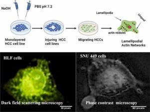
The migratory process is a highly organized, differentiated, and polarized stage by which many signaling pathways are regulated to control cell migration. Since the significant evidence of migrating cells is the reorganization of the cytoskeleton. In the recent study, the cell migration model was assessed on the fact that any disruption obtained in the cellular monolayer confluent, may cause stimulation for surrounding cells to migrate. We attempt to demonstrate the morphological alterations associated with these migrating cells. In this case, sterilized 1 N NaOH (1 µl) was used as alkaline burnt. It leads to scratching the monolayer of hepatocellular carcinoma (HLF cell line) allowing cells to lose their connection. Scanning electron microscopy (SEM), fluorescence microscopy, light inverted microscopy, and dark field were used for discovering the morphological alterations associated with migrating cancer cells. The findings show that cells exhibited distinctive alterations including a polarizing stage, accumulation of the actin nodules in front of the nucleus, and protrusions. Nuclei appeared as lobulated shapes during migration. Lamellipodia and uropod were extended as well. Additionally, TGFβ1 proved its expression in HLF and SNU449 after their stimulation. It is demonstrated that hepatocellular carcinoma cells can migrate after their stimulation and there is a caution against the indiscriminate application of alkalinizing drug therapy.
期刊介绍:
Acta histochemica, a journal of structural biochemistry of cells and tissues, publishes original research articles, short communications, reviews, letters to the editor, meeting reports and abstracts of meetings. The aim of the journal is to provide a forum for the cytochemical and histochemical research community in the life sciences, including cell biology, biotechnology, neurobiology, immunobiology, pathology, pharmacology, botany, zoology and environmental and toxicological research. The journal focuses on new developments in cytochemistry and histochemistry and their applications. Manuscripts reporting on studies of living cells and tissues are particularly welcome. Understanding the complexity of cells and tissues, i.e. their biocomplexity and biodiversity, is a major goal of the journal and reports on this topic are especially encouraged. Original research articles, short communications and reviews that report on new developments in cytochemistry and histochemistry are welcomed, especially when molecular biology is combined with the use of advanced microscopical techniques including image analysis and cytometry. Letters to the editor should comment or interpret previously published articles in the journal to trigger scientific discussions. Meeting reports are considered to be very important publications in the journal because they are excellent opportunities to present state-of-the-art overviews of fields in research where the developments are fast and hard to follow. Authors of meeting reports should consult the editors before writing a report. The editorial policy of the editors and the editorial board is rapid publication. Once a manuscript is received by one of the editors, an editorial decision about acceptance, revision or rejection will be taken within a month. It is the aim of the publishers to have a manuscript published within three months after the manuscript has been accepted


 分享
分享
 求助内容:
求助内容: 应助结果提醒方式:
 扫码关注我们
