{"title":"Does sevoflurane sedation in pediatric patients lead to \"pseudo\" leptomeningeal enhancement in the brain on 3 Tesla magnetic resonance imaging?","authors":"Kiran Hilal, Kumail Khandwala, Saima Rashid, Faheemullah Khan, Shayan Sirat Maheen Anwar","doi":"10.4329/wjr.v15.i4.127","DOIUrl":null,"url":null,"abstract":"<p><strong>Background: </strong>Prominent leptomeningeal contrast enhancement (LMCE) in the brain is observed in some pediatric patients during sedation for imaging. However, based on clinical history and cerebrospinal fluid analysis, the patients are not acutely ill and do not exhibit meningeal signs. Our study determined whether sevoflurane inhalation in pediatric patients led to this pattern of 'pseudo' LMCE (pLMCE) on 3 Tesla magnetic resonance imaging (MRI).</p><p><strong>Aim: </strong>To highlight the significance of pLMCE in pediatric patients undergoing enhanced brain MRI under sedation to avoid misinterpretation in reports.</p><p><strong>Methods: </strong>A retrospective cross-sectional evaluation of pediatric patients between 0-8 years of age was conducted. The patients underwent enhanced brain MRI under inhaled sevoflurane. The LMCE grade was determined by two radiologists, and interobserver variability of the grade was calculated using Cohen's kappa. The LMCE grade was correlated with duration of sedation, age and weight using the Spearman rho rank correlation.</p><p><strong>Results: </strong>A total of 63 patients were included. Fourteen (22.2%) cases showed mild LMCE, 48 (76.1%) cases showed moderate LMCE, and 1 case (1.6%) showed severe LMCE. We found substantial agreement between the two radiologists in detection of pLMCE on post-contrast T1 imaging (kappa value = 0.61; <i>P</i> < 0.001). Additionally, we found statistically significant inverse and moderate correlations between patient weight and age. There was no correlation between duration of sedation and pLMCE.</p><p><strong>Conclusion: </strong>pLMCE is relatively common on post-contrast spin echo T1-weighted MRI of pediatric patients sedated by sevoflurane due to their fragile and immature vasculature. It should not be misinterpreted for meningeal pathology. Knowing pertinent clinical history of the child is an essential prerequisite to avoid radiological overcalling and the subsequent burden of additional investigations.</p>","PeriodicalId":23819,"journal":{"name":"World journal of radiology","volume":"15 4","pages":"127-135"},"PeriodicalIF":1.5000,"publicationDate":"2023-04-28","publicationTypes":"Journal Article","fieldsOfStudy":null,"isOpenAccess":false,"openAccessPdf":"https://ftp.ncbi.nlm.nih.gov/pub/pmc/oa_pdf/79/97/WJR-15-127.PMC10167815.pdf","citationCount":"0","resultStr":null,"platform":"Semanticscholar","paperid":null,"PeriodicalName":"World journal of radiology","FirstCategoryId":"1085","ListUrlMain":"https://doi.org/10.4329/wjr.v15.i4.127","RegionNum":0,"RegionCategory":null,"ArticlePicture":[],"TitleCN":null,"AbstractTextCN":null,"PMCID":null,"EPubDate":"","PubModel":"","JCR":"Q3","JCRName":"RADIOLOGY, NUCLEAR MEDICINE & MEDICAL IMAGING","Score":null,"Total":0}
引用次数: 0
Abstract
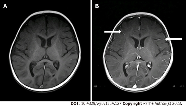
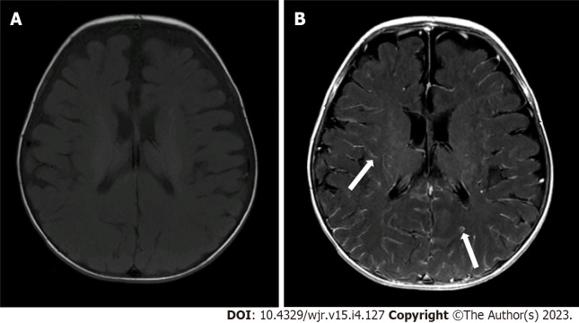
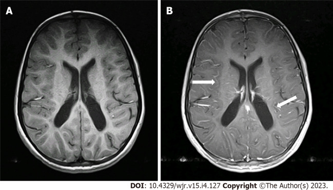
Background: Prominent leptomeningeal contrast enhancement (LMCE) in the brain is observed in some pediatric patients during sedation for imaging. However, based on clinical history and cerebrospinal fluid analysis, the patients are not acutely ill and do not exhibit meningeal signs. Our study determined whether sevoflurane inhalation in pediatric patients led to this pattern of 'pseudo' LMCE (pLMCE) on 3 Tesla magnetic resonance imaging (MRI).
Aim: To highlight the significance of pLMCE in pediatric patients undergoing enhanced brain MRI under sedation to avoid misinterpretation in reports.
Methods: A retrospective cross-sectional evaluation of pediatric patients between 0-8 years of age was conducted. The patients underwent enhanced brain MRI under inhaled sevoflurane. The LMCE grade was determined by two radiologists, and interobserver variability of the grade was calculated using Cohen's kappa. The LMCE grade was correlated with duration of sedation, age and weight using the Spearman rho rank correlation.
Results: A total of 63 patients were included. Fourteen (22.2%) cases showed mild LMCE, 48 (76.1%) cases showed moderate LMCE, and 1 case (1.6%) showed severe LMCE. We found substantial agreement between the two radiologists in detection of pLMCE on post-contrast T1 imaging (kappa value = 0.61; P < 0.001). Additionally, we found statistically significant inverse and moderate correlations between patient weight and age. There was no correlation between duration of sedation and pLMCE.
Conclusion: pLMCE is relatively common on post-contrast spin echo T1-weighted MRI of pediatric patients sedated by sevoflurane due to their fragile and immature vasculature. It should not be misinterpreted for meningeal pathology. Knowing pertinent clinical history of the child is an essential prerequisite to avoid radiological overcalling and the subsequent burden of additional investigations.


 分享
分享
 求助内容:
求助内容: 应助结果提醒方式:
 扫码关注我们
