Çağdaş Özgökçe, Aydın Öcal, Işılay Seze Ermiş, Engin Deveci
{"title":"Histopathological, ultrastructural, and immunohistochemical examination of changes in the placenta as a result of severe preeclampsia.","authors":"Çağdaş Özgökçe, Aydın Öcal, Işılay Seze Ermiş, Engin Deveci","doi":"10.1590/acb382023","DOIUrl":null,"url":null,"abstract":"<p><strong>Purpose: </strong>To investigate the role of hypoxia-inducible transcription factor-1 alpha (HIF-1α) and angiogenetic factor endothelin-1 (ET-1) expression in regulating hypoxia and placental development by routine histopathological methods.</p><p><strong>Methods: </strong>Twenty preeclamptic and normal placentas were used. Placenta tissue pieces were examined histopathologically after routine paraffin follow-ups. HIF-1α and ET-1 proteins were examined immunohistochemically, and placental tissues were examined ultrastructurally.</p><p><strong>Results: </strong>Increase in syncytial proliferation, endothelial damage in vessels, and increase in collagen were observed in preeclamptic placentas. As a result of preeclampsia, an increase was observed in HIF-1α and ET-1 protein levels in the placenta. Dilatation of endoplasmic reticulum and loss of cristae in mitochondria were observed in trophoblast cells in preeclamptic placental sections.</p><p><strong>Conclusions: </strong>High regulation of oxygen resulting from preeclampsia has been shown to be a critical determinant of placentagenesis and plays an important role in placental differentiation, changes in maternal and fetal blood circulation, trophoblastic invasion, and syncytial node increase. It has been thought that preeclampsia affects secretion by disrupting the endoplasmic reticulum structure and induces mitochondrial damage, and that ET-1 may potentially help in the induction of stress pathways as a result of hypoxia in preeclampsia.</p>","PeriodicalId":6992,"journal":{"name":"Acta cirurgica brasileira","volume":"38 ","pages":"e382023"},"PeriodicalIF":1.3000,"publicationDate":"2023-01-01","publicationTypes":"Journal Article","fieldsOfStudy":null,"isOpenAccess":false,"openAccessPdf":"https://www.ncbi.nlm.nih.gov/pmc/articles/PMC10191158/pdf/","citationCount":"0","resultStr":null,"platform":"Semanticscholar","paperid":null,"PeriodicalName":"Acta cirurgica brasileira","FirstCategoryId":"3","ListUrlMain":"https://doi.org/10.1590/acb382023","RegionNum":4,"RegionCategory":"医学","ArticlePicture":[],"TitleCN":null,"AbstractTextCN":null,"PMCID":null,"EPubDate":"","PubModel":"","JCR":"Q3","JCRName":"SURGERY","Score":null,"Total":0}
引用次数: 0
Abstract
Purpose: To investigate the role of hypoxia-inducible transcription factor-1 alpha (HIF-1α) and angiogenetic factor endothelin-1 (ET-1) expression in regulating hypoxia and placental development by routine histopathological methods.
Methods: Twenty preeclamptic and normal placentas were used. Placenta tissue pieces were examined histopathologically after routine paraffin follow-ups. HIF-1α and ET-1 proteins were examined immunohistochemically, and placental tissues were examined ultrastructurally.
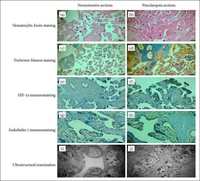
Results: Increase in syncytial proliferation, endothelial damage in vessels, and increase in collagen were observed in preeclamptic placentas. As a result of preeclampsia, an increase was observed in HIF-1α and ET-1 protein levels in the placenta. Dilatation of endoplasmic reticulum and loss of cristae in mitochondria were observed in trophoblast cells in preeclamptic placental sections.
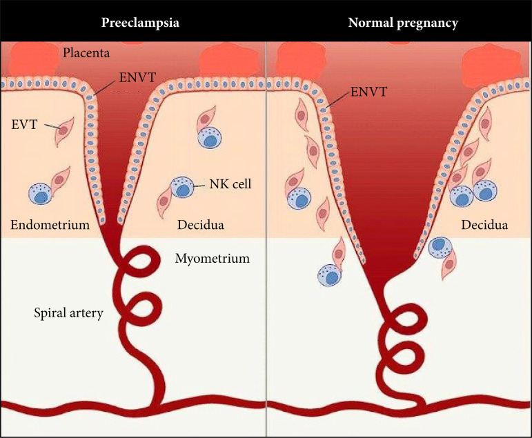
Conclusions: High regulation of oxygen resulting from preeclampsia has been shown to be a critical determinant of placentagenesis and plays an important role in placental differentiation, changes in maternal and fetal blood circulation, trophoblastic invasion, and syncytial node increase. It has been thought that preeclampsia affects secretion by disrupting the endoplasmic reticulum structure and induces mitochondrial damage, and that ET-1 may potentially help in the induction of stress pathways as a result of hypoxia in preeclampsia.


 分享
分享
 求助内容:
求助内容: 应助结果提醒方式:
 扫码关注我们
