Michaela Aurelia Maria Huson, Tapiwa Kumwenda, Joe Gumulira, Ethel Rambiki, Claudia Wallrauch, Tom Heller
{"title":"Ultrasound findings in Kaposi sarcoma patients: overlapping sonographic features with disseminated tuberculosis.","authors":"Michaela Aurelia Maria Huson, Tapiwa Kumwenda, Joe Gumulira, Ethel Rambiki, Claudia Wallrauch, Tom Heller","doi":"10.1186/s13089-023-00323-8","DOIUrl":null,"url":null,"abstract":"<p><strong>Background: </strong>Focused Assessment with Sonography for HIV-associated TB (FASH) is a diagnostic tool for extra-pulmonary tuberculosis (TB) in symptomatic patients with advanced HIV. As Kaposi's sarcoma (KS) is also prevalent in this patient population, changes due to KS may mimic TB findings and clinical interpretation of target FASH findings can be challenging. We aimed to describe sonographic findings in patients with KS.</p><p><strong>Methods: </strong>We performed a prospective observational study at Lighthouse clinic at Kamuzu Central Hospital, Lilongwe, Malawi, in consecutive patients with newly diagnosed KS, without known diagnosis of TB, referred for paclitaxel treatment. All patients underwent FASH and abdominal ultrasound to assess for effusions and changes in liver and spleen, as well as systematic sonographic assessment for lymphadenopathy.</p><p><strong>Results: </strong>We included 30 patients. We found inguinal lymph nodes using ultrasound in 20 patients; in 3 (10%) additionally abdominal lymph nodes were found. Pathological effusions were seen in eight patients (27%): pericardial effusion in one (3%), pleural effusion in six (20%) and ascites in four (13%) patients. We found focal spleen lesions in three (10%) patients. Most of these lesions were echogenic, but in one patient, we saw hypoechoic lesions with an echogenic center. In three (10%) patients an unusual \"sponge-like pattern\" of the splenic vasculature was found. Six (20%) patients had echogenic focal lesions in the liver resembling hemangiomas, individual lesions showing a hypoechoic center. In two patients echogenic portal fields were seen.</p><p><strong>Conclusions: </strong>The majority of patients with newly diagnosed KS demonstrate sonographic features of disease, predominantly lymphadenopathy. Effusions were observed in a significant minority, as well as focal lesions in liver or spleen, which commonly resemble hemangiomas, but hypoechoic lesions were also observed and can easily be mistaken for extra-pulmonary TB. A 'sponge-like pattern' of the spleen should not be confused with micro-abscesses. In conclusion, this case series illustrates the diverse nature of ultrasound features in patients with KS, which can be difficult to distinguish from other opportunistic diseases, including TB.</p>","PeriodicalId":75201,"journal":{"name":"","volume":"15 1","pages":"27"},"PeriodicalIF":0.0,"publicationDate":"2023-06-01","publicationTypes":"Journal Article","fieldsOfStudy":null,"isOpenAccess":false,"openAccessPdf":"https://www.ncbi.nlm.nih.gov/pmc/articles/PMC10232376/pdf/","citationCount":"2","resultStr":null,"platform":"Semanticscholar","paperid":null,"PeriodicalName":"","FirstCategoryId":"1085","ListUrlMain":"https://doi.org/10.1186/s13089-023-00323-8","RegionNum":0,"RegionCategory":null,"ArticlePicture":[],"TitleCN":null,"AbstractTextCN":null,"PMCID":null,"EPubDate":"","PubModel":"","JCR":"","JCRName":"","Score":null,"Total":0}
引用次数: 2
Abstract
Background: Focused Assessment with Sonography for HIV-associated TB (FASH) is a diagnostic tool for extra-pulmonary tuberculosis (TB) in symptomatic patients with advanced HIV. As Kaposi's sarcoma (KS) is also prevalent in this patient population, changes due to KS may mimic TB findings and clinical interpretation of target FASH findings can be challenging. We aimed to describe sonographic findings in patients with KS.
Methods: We performed a prospective observational study at Lighthouse clinic at Kamuzu Central Hospital, Lilongwe, Malawi, in consecutive patients with newly diagnosed KS, without known diagnosis of TB, referred for paclitaxel treatment. All patients underwent FASH and abdominal ultrasound to assess for effusions and changes in liver and spleen, as well as systematic sonographic assessment for lymphadenopathy.
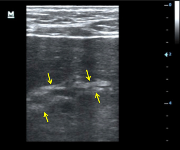
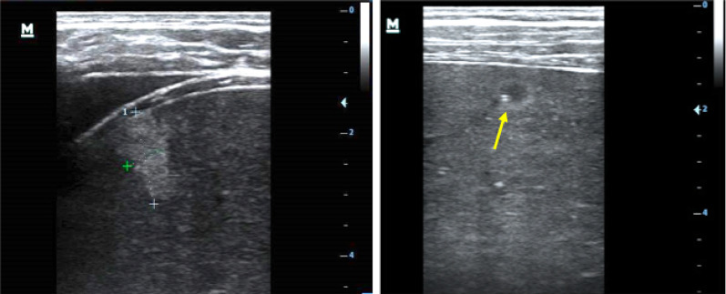
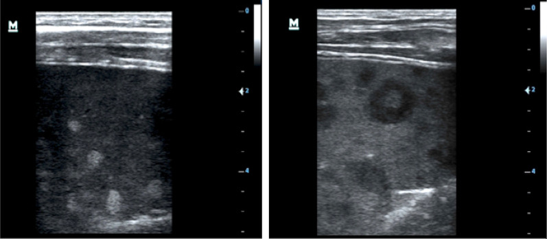
Results: We included 30 patients. We found inguinal lymph nodes using ultrasound in 20 patients; in 3 (10%) additionally abdominal lymph nodes were found. Pathological effusions were seen in eight patients (27%): pericardial effusion in one (3%), pleural effusion in six (20%) and ascites in four (13%) patients. We found focal spleen lesions in three (10%) patients. Most of these lesions were echogenic, but in one patient, we saw hypoechoic lesions with an echogenic center. In three (10%) patients an unusual "sponge-like pattern" of the splenic vasculature was found. Six (20%) patients had echogenic focal lesions in the liver resembling hemangiomas, individual lesions showing a hypoechoic center. In two patients echogenic portal fields were seen.
Conclusions: The majority of patients with newly diagnosed KS demonstrate sonographic features of disease, predominantly lymphadenopathy. Effusions were observed in a significant minority, as well as focal lesions in liver or spleen, which commonly resemble hemangiomas, but hypoechoic lesions were also observed and can easily be mistaken for extra-pulmonary TB. A 'sponge-like pattern' of the spleen should not be confused with micro-abscesses. In conclusion, this case series illustrates the diverse nature of ultrasound features in patients with KS, which can be difficult to distinguish from other opportunistic diseases, including TB.


 分享
分享
 求助内容:
求助内容: 应助结果提醒方式:
 扫码关注我们
