{"title":"Effects of allium cepa on ovarian torsion-detorsion injury in a rat model.","authors":"Hakan Kula, Orkun İlgen, Sefa Kurt, Filiz Yılmaz","doi":"10.4274/tjod.galenos.2023.41763","DOIUrl":null,"url":null,"abstract":"<p><strong>Objective: </strong>Ischemia/reperfusion (I/R) damage following detorsion treatment, tissue fibrosis, and adhesions cause secondary tissue damage in the ovaries. Many studies have been evaluated to minimize antioxidant damage in ovarian reserve loss while minimizing I/R damage. However, no study observed long-term effects on the ovarian torsion model in rats. In this study, we evaluated the profibrotic effects of A. cepa on an ovarian torsion model on rats.</p><p><strong>Materials and methods: </strong>Group I (n=7) rats were the sham group. Group II (n=7) rats were the torsion group and Group III (n=7) rats were the torsion + A. cepa group. To observe the long-term effects of allium cepa, rats were fed for 21 days. Cellular damage I/R is evaluated by histopathological damage score, and transforming growth factor-beta 1 (TGF-β1) and alpha-smooth muscle actin (α-SMA) is measured to analyze the profibrotic effect.</p><p><strong>Results: </strong>A. cepa altered cellular damage due to improvement in the histopathological damage score with A. cepa intake. However, the profibrotic mediators TGF-β1 and α-SMA are non- significantly changed by the A. cepa (p=0.477 and p=0.185 respectively).</p><p><strong>Conclusion: </strong>A. cepa is a potent protective on cellular tissue, minimizing I/R damage on ovarian tissue histologically. Our study implies that A. cepa does not affect fibrosis-related mediators in the rat ovary.</p>","PeriodicalId":45340,"journal":{"name":"Turkish Journal of Obstetrics and Gynecology","volume":"20 2","pages":"137-141"},"PeriodicalIF":1.3000,"publicationDate":"2023-06-01","publicationTypes":"Journal Article","fieldsOfStudy":null,"isOpenAccess":false,"openAccessPdf":"https://ftp.ncbi.nlm.nih.gov/pub/pmc/oa_pdf/6e/3a/TJOG-20-137.PMC10236230.pdf","citationCount":"0","resultStr":null,"platform":"Semanticscholar","paperid":null,"PeriodicalName":"Turkish Journal of Obstetrics and Gynecology","FirstCategoryId":"1085","ListUrlMain":"https://doi.org/10.4274/tjod.galenos.2023.41763","RegionNum":0,"RegionCategory":null,"ArticlePicture":[],"TitleCN":null,"AbstractTextCN":null,"PMCID":null,"EPubDate":"","PubModel":"","JCR":"Q4","JCRName":"OBSTETRICS & GYNECOLOGY","Score":null,"Total":0}
引用次数: 0
Abstract
Objective: Ischemia/reperfusion (I/R) damage following detorsion treatment, tissue fibrosis, and adhesions cause secondary tissue damage in the ovaries. Many studies have been evaluated to minimize antioxidant damage in ovarian reserve loss while minimizing I/R damage. However, no study observed long-term effects on the ovarian torsion model in rats. In this study, we evaluated the profibrotic effects of A. cepa on an ovarian torsion model on rats.
Materials and methods: Group I (n=7) rats were the sham group. Group II (n=7) rats were the torsion group and Group III (n=7) rats were the torsion + A. cepa group. To observe the long-term effects of allium cepa, rats were fed for 21 days. Cellular damage I/R is evaluated by histopathological damage score, and transforming growth factor-beta 1 (TGF-β1) and alpha-smooth muscle actin (α-SMA) is measured to analyze the profibrotic effect.
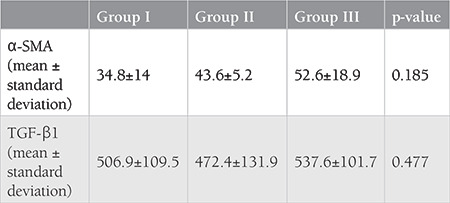
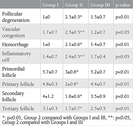
Results: A. cepa altered cellular damage due to improvement in the histopathological damage score with A. cepa intake. However, the profibrotic mediators TGF-β1 and α-SMA are non- significantly changed by the A. cepa (p=0.477 and p=0.185 respectively).
Conclusion: A. cepa is a potent protective on cellular tissue, minimizing I/R damage on ovarian tissue histologically. Our study implies that A. cepa does not affect fibrosis-related mediators in the rat ovary.


 分享
分享
 求助内容:
求助内容: 应助结果提醒方式:
 扫码关注我们
