{"title":"Transfer learning in diagnosis of maxillary sinusitis using panoramic radiography and conventional radiography.","authors":"Shinya Kotaki, Takahito Nishiguchi, Marino Araragi, Hironori Akiyama, Motoki Fukuda, Eiichiro Ariji, Yoshiko Ariji","doi":"10.1007/s11282-022-00658-3","DOIUrl":null,"url":null,"abstract":"<p><strong>Objectives: </strong>To clarify the performance of transfer learning with a small number of Waters' images at institution B in diagnosing maxillary sinusitis, based on a source model trained with a large number of panoramic radiographs at institution A.</p><p><strong>Methods: </strong>The source model was created by a 200-epoch training process with 800 training and 60 validation datasets of panoramic radiographs at institution A using VGG-16. One hundred and eighty Waters' and 180 panoramic image patches with or without maxillary sinusitis at institution B were enrolled in this study, and were arbitrarily assigned to 120 training, 20 validation, and 40 test datasets, respectively. Transfer learning of 200 epochs was performed using the training and validation datasets of Waters' images based on the source model, and the target model was obtained. The test Waters' images were applied to the source and target models, and the performance of each model was evaluated. Transfer learning with panoramic radiographs and evaluation by two radiologists were undertaken and compared. The evaluation was based on the area of receiver-operating characteristic curves (AUC).</p><p><strong>Results: </strong>When using Waters' images as the test dataset, the AUCs of the source model, target model, and radiologists were 0.780, 0.830, and 0.806, respectively. There were no significant differences between these models and the radiologists, whereas the target model performed better than the source model. For panoramic radiographs, AUCs were 0.863, 0.863, and 0.808, respectively, with no significant differences.</p><p><strong>Conclusions: </strong>This study performed transfer learning using a small number of Waters' images, based on a source model created solely from panoramic radiographs, resulting in a performance improvement to 0.830 in diagnosing maxillary sinusitis, which was equivalent to that of radiologists. Transfer learning is considered a useful method to improve diagnostic performance.</p>","PeriodicalId":56103,"journal":{"name":"Oral Radiology","volume":"39 3","pages":"467-474"},"PeriodicalIF":1.7000,"publicationDate":"2023-07-01","publicationTypes":"Journal Article","fieldsOfStudy":null,"isOpenAccess":false,"openAccessPdf":"","citationCount":"0","resultStr":null,"platform":"Semanticscholar","paperid":null,"PeriodicalName":"Oral Radiology","FirstCategoryId":"3","ListUrlMain":"https://doi.org/10.1007/s11282-022-00658-3","RegionNum":3,"RegionCategory":"医学","ArticlePicture":[],"TitleCN":null,"AbstractTextCN":null,"PMCID":null,"EPubDate":"","PubModel":"","JCR":"Q3","JCRName":"DENTISTRY, ORAL SURGERY & MEDICINE","Score":null,"Total":0}
引用次数: 0
Abstract
Objectives: To clarify the performance of transfer learning with a small number of Waters' images at institution B in diagnosing maxillary sinusitis, based on a source model trained with a large number of panoramic radiographs at institution A.
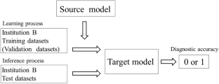
Methods: The source model was created by a 200-epoch training process with 800 training and 60 validation datasets of panoramic radiographs at institution A using VGG-16. One hundred and eighty Waters' and 180 panoramic image patches with or without maxillary sinusitis at institution B were enrolled in this study, and were arbitrarily assigned to 120 training, 20 validation, and 40 test datasets, respectively. Transfer learning of 200 epochs was performed using the training and validation datasets of Waters' images based on the source model, and the target model was obtained. The test Waters' images were applied to the source and target models, and the performance of each model was evaluated. Transfer learning with panoramic radiographs and evaluation by two radiologists were undertaken and compared. The evaluation was based on the area of receiver-operating characteristic curves (AUC).
Results: When using Waters' images as the test dataset, the AUCs of the source model, target model, and radiologists were 0.780, 0.830, and 0.806, respectively. There were no significant differences between these models and the radiologists, whereas the target model performed better than the source model. For panoramic radiographs, AUCs were 0.863, 0.863, and 0.808, respectively, with no significant differences.
Conclusions: This study performed transfer learning using a small number of Waters' images, based on a source model created solely from panoramic radiographs, resulting in a performance improvement to 0.830 in diagnosing maxillary sinusitis, which was equivalent to that of radiologists. Transfer learning is considered a useful method to improve diagnostic performance.
期刊介绍:
As the official English-language journal of the Japanese Society for Oral and Maxillofacial Radiology and the Asian Academy of Oral and Maxillofacial Radiology, Oral Radiology is intended to be a forum for international collaboration in head and neck diagnostic imaging and all related fields. Oral Radiology features cutting-edge research papers, review articles, case reports, and technical notes from both the clinical and experimental fields. As membership in the Society is not a prerequisite, contributions are welcome from researchers and clinicians worldwide.


 分享
分享
 求助内容:
求助内容: 应助结果提醒方式:
 扫码关注我们
