Boxin Xue, Caiwei Zhou, Yizhi Qin, Yongzheng Li, Yuao Sun, Lei Chang, Shipeng Shao, Yongliang Li, Mengling Zhang, Chaoying Sun, Renxi He, Qian Peter Su, Yujie Sun
{"title":"PN-ImTLSM facilitates high-throughput low background single-molecule localization microscopy deep in the cell.","authors":"Boxin Xue, Caiwei Zhou, Yizhi Qin, Yongzheng Li, Yuao Sun, Lei Chang, Shipeng Shao, Yongliang Li, Mengling Zhang, Chaoying Sun, Renxi He, Qian Peter Su, Yujie Sun","doi":"10.52601/bpr.2021.210014","DOIUrl":null,"url":null,"abstract":"<p><p>When imaging the nucleus structure of a cell, the out-of-focus fluorescence acts as background and hinders the detection of weak signals. Light-sheet fluorescence microscopy (LSFM) is a wide-field imaging approach which has the best of both background removal and imaging speed. However, the commonly adopted orthogonal excitation/detection scheme is hard to be applied to single-cell imaging due to steric hindrance. For LSFMs capable of high spatiotemporal single-cell imaging, the complex instrument design and operation largely limit their throughput of data collection. Here, we propose an approach for high-throughput background-free fluorescence imaging of single cells facilitated by the Immersion Tilted Light Sheet Microscopy (ImTLSM). ImTLSM is based on a light-sheet projected off the optical axis of a water immersion objective. With the illumination objective and the detection objective placed opposingly, ImTLSM can rapidly patrol and optically section multiple individual cells while maintaining single-molecule detection sensitivity and resolution. Further, the simplicity and robustness of ImTLSM in operation and maintenance enables high-throughput image collection to establish background removal datasets for deep learning. Using a deep learning model to train the mapping from epi-illumination images to ImTLSM illumination images, namely PN-ImTLSM, we demonstrated cross-modality fluorescence imaging, transforming the epi-illumination image to approach the background removal performance obtained with ImTLSM. We demonstrated that PN-ImTLSM can be generalized to large-field homogeneous illumination imaging, thereby further improving the imaging throughput. In addition, compared to commonly used background removal methods, PN-ImTLSM showed much better performance for areas where the background intensity changes sharply in space, facilitating high-density single-molecule localization microscopy. In summary, PN-ImTLSM paves the way for background-free fluorescence imaging on ordinary inverted microscopes.</p>","PeriodicalId":59621,"journal":{"name":"生物物理学报:英文版","volume":"7 4","pages":"313-325"},"PeriodicalIF":0.0000,"publicationDate":"2021-08-31","publicationTypes":"Journal Article","fieldsOfStudy":null,"isOpenAccess":false,"openAccessPdf":"https://www.ncbi.nlm.nih.gov/pmc/articles/PMC10233473/pdf/","citationCount":"0","resultStr":null,"platform":"Semanticscholar","paperid":null,"PeriodicalName":"生物物理学报:英文版","FirstCategoryId":"1085","ListUrlMain":"https://doi.org/10.52601/bpr.2021.210014","RegionNum":0,"RegionCategory":null,"ArticlePicture":[],"TitleCN":null,"AbstractTextCN":null,"PMCID":null,"EPubDate":"","PubModel":"","JCR":"","JCRName":"","Score":null,"Total":0}
引用次数: 0
Abstract
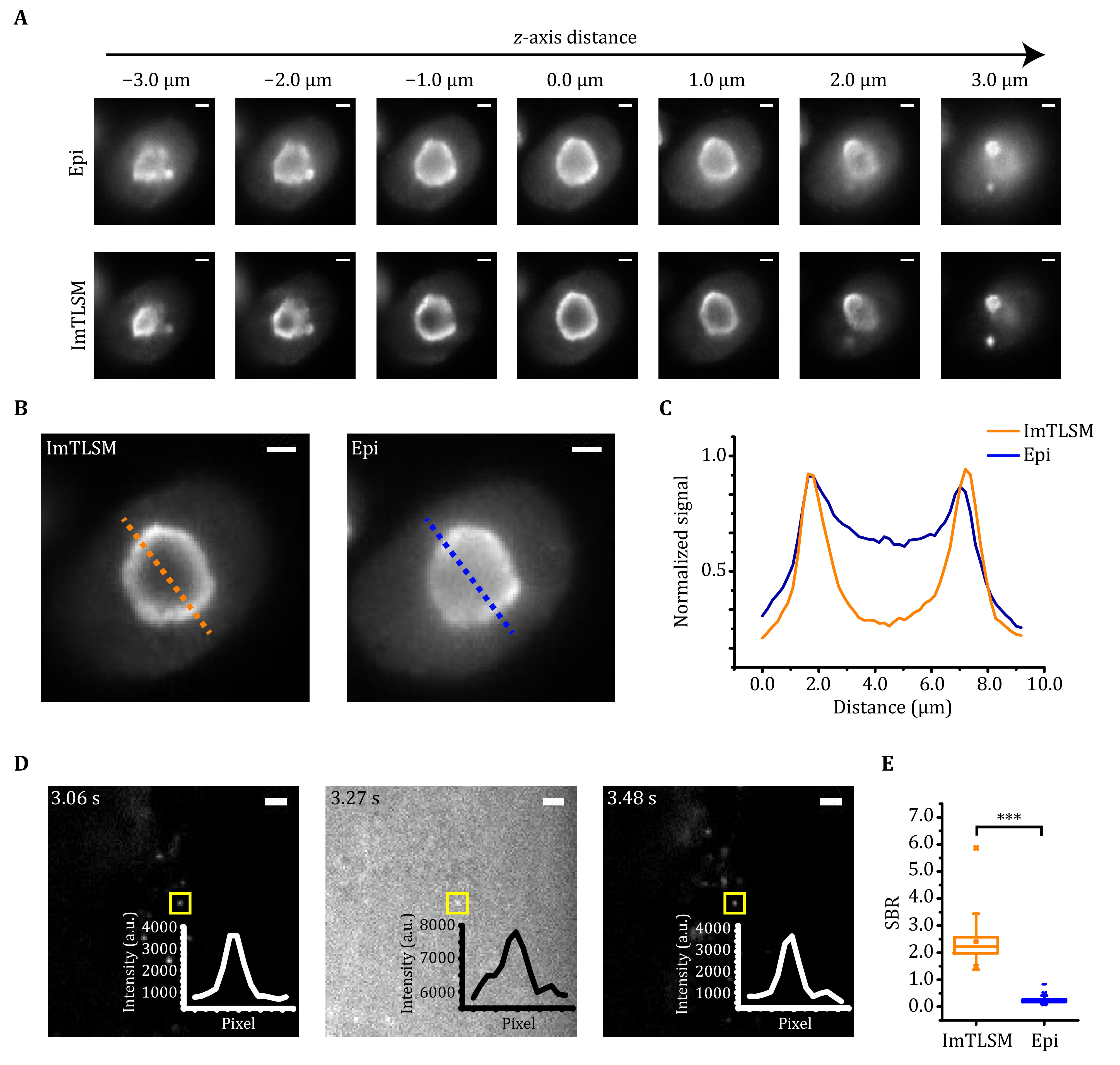
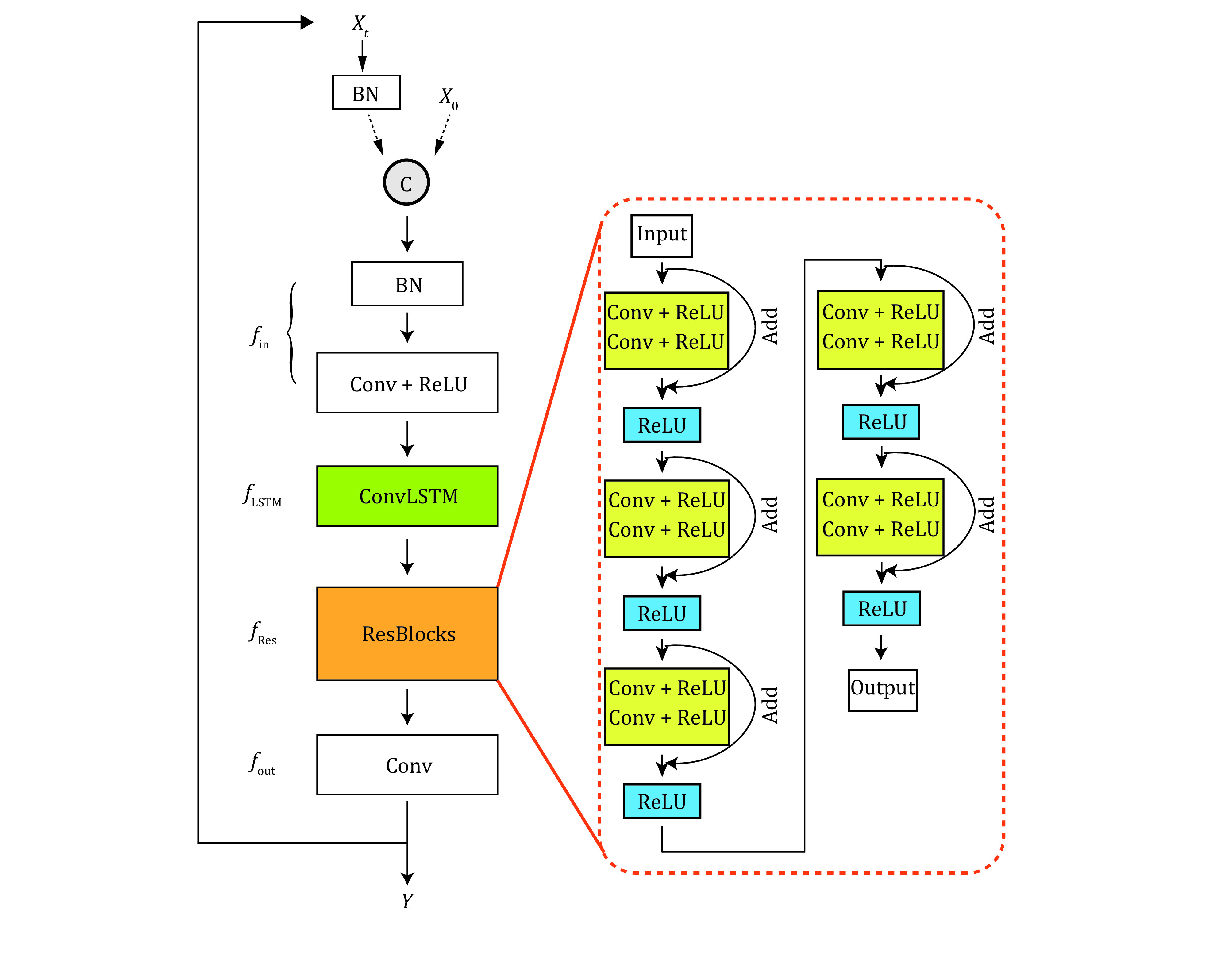
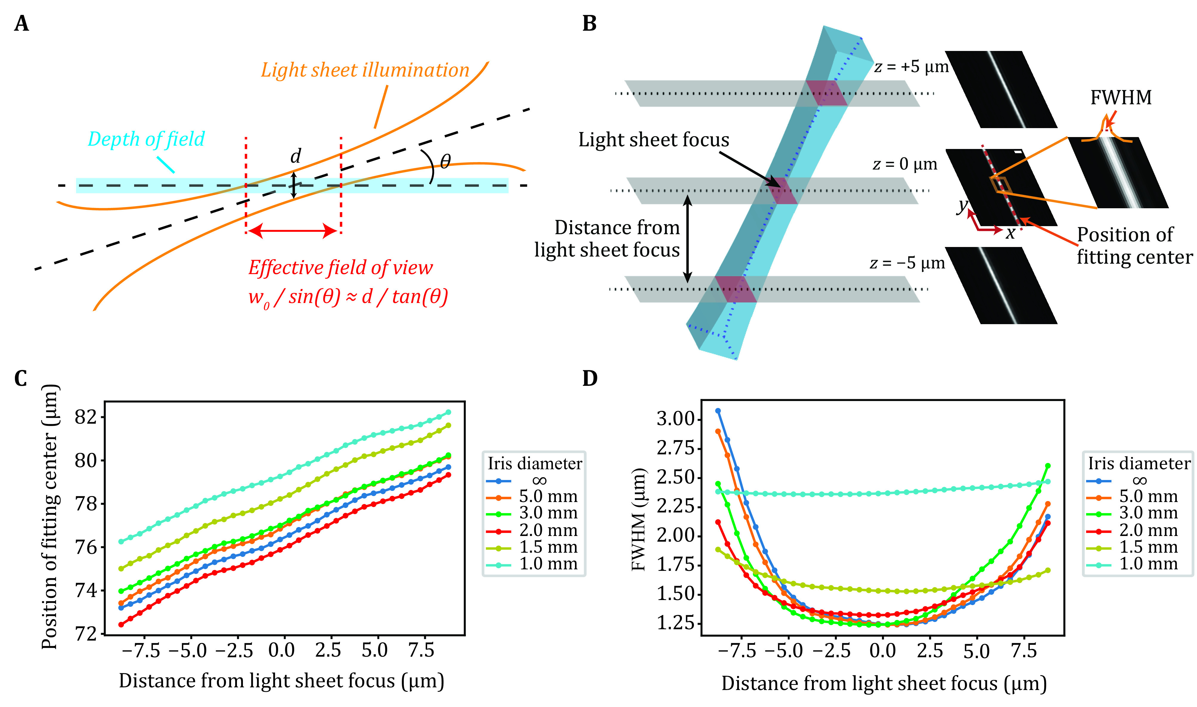
When imaging the nucleus structure of a cell, the out-of-focus fluorescence acts as background and hinders the detection of weak signals. Light-sheet fluorescence microscopy (LSFM) is a wide-field imaging approach which has the best of both background removal and imaging speed. However, the commonly adopted orthogonal excitation/detection scheme is hard to be applied to single-cell imaging due to steric hindrance. For LSFMs capable of high spatiotemporal single-cell imaging, the complex instrument design and operation largely limit their throughput of data collection. Here, we propose an approach for high-throughput background-free fluorescence imaging of single cells facilitated by the Immersion Tilted Light Sheet Microscopy (ImTLSM). ImTLSM is based on a light-sheet projected off the optical axis of a water immersion objective. With the illumination objective and the detection objective placed opposingly, ImTLSM can rapidly patrol and optically section multiple individual cells while maintaining single-molecule detection sensitivity and resolution. Further, the simplicity and robustness of ImTLSM in operation and maintenance enables high-throughput image collection to establish background removal datasets for deep learning. Using a deep learning model to train the mapping from epi-illumination images to ImTLSM illumination images, namely PN-ImTLSM, we demonstrated cross-modality fluorescence imaging, transforming the epi-illumination image to approach the background removal performance obtained with ImTLSM. We demonstrated that PN-ImTLSM can be generalized to large-field homogeneous illumination imaging, thereby further improving the imaging throughput. In addition, compared to commonly used background removal methods, PN-ImTLSM showed much better performance for areas where the background intensity changes sharply in space, facilitating high-density single-molecule localization microscopy. In summary, PN-ImTLSM paves the way for background-free fluorescence imaging on ordinary inverted microscopes.


 分享
分享

 求助内容:
求助内容: 应助结果提醒方式:
 扫码关注我们
