{"title":"Visualizing the triheteromeric N-methyl-D-aspartate receptor subunit composition.","authors":"Stephen Beesley, Akash Gunjan, Sanjay S Kumar","doi":"10.3389/fnsyn.2023.1156777","DOIUrl":null,"url":null,"abstract":"<p><p>N-methyl-D-aspartate receptors (NMDARs) are one of three ligand-gated ionotropic channels that transduce the effects of neurotransmitter glutamate at excitatory synapses within the central nervous system. Their ability to influx Ca<sup>2+</sup> into cells, unlike mature AMPA or kainate receptors, implicates them in a variety of processes ranging from synaptic plasticity to cell death. Many of the receptor's capabilities, including binding glutamate and regulating Ca<sup>2+</sup> influx, have been attributed to their subunit composition, determined putatively using cell biology, electrophysiology and/or pharmacology. Here, we show that subunit composition of synaptic NMDARs can also be readily visualized in acute brain slices (rat) using highly specific antibodies directed against extracellular epitopes of the subunit proteins and high-resolution confocal microscopy. This has helped confirm the expression of triheteromeric <i>t</i>-NMDARs (containing GluN1, GluN2, and GluN3 subunits) at synapses for the first time and reconcile functional differences with diheteromeric <i>d</i>-NMDARs (containing GluN1 and GluN2 subunits) described previously. Even though structural information about individual receptors is still diffraction limited, fluorescently tagged receptor subunit puncta coalesce with precision at various magnifications and/or with the postsynaptic density (PSD-95) but not the presynaptic active zone marker Bassoon. These data are particularly relevant for identifying GluN3A-containing <i>t</i>-NMDARs that are highly Ca<sup>2+</sup> permeable and whose expression at excitatory synapses renders neurons vulnerable to excitotoxicity and cell death. Imaging NMDAR subunit proteins at synapses not only offers firsthand insights into subunit composition to correlate function but may also help identify zones of vulnerability within brain structures underlying neurodegenerative diseases like Temporal Lobe Epilepsy.</p>","PeriodicalId":12650,"journal":{"name":"Frontiers in Synaptic Neuroscience","volume":"15 ","pages":"1156777"},"PeriodicalIF":4.1000,"publicationDate":"2023-05-24","publicationTypes":"Journal Article","fieldsOfStudy":null,"isOpenAccess":false,"openAccessPdf":"https://www.ncbi.nlm.nih.gov/pmc/articles/PMC10244591/pdf/","citationCount":"0","resultStr":null,"platform":"Semanticscholar","paperid":null,"PeriodicalName":"Frontiers in Synaptic Neuroscience","FirstCategoryId":"3","ListUrlMain":"https://doi.org/10.3389/fnsyn.2023.1156777","RegionNum":4,"RegionCategory":"医学","ArticlePicture":[],"TitleCN":null,"AbstractTextCN":null,"PMCID":null,"EPubDate":"2023/1/1 0:00:00","PubModel":"eCollection","JCR":"Q2","JCRName":"NEUROSCIENCES","Score":null,"Total":0}
引用次数: 0
Abstract
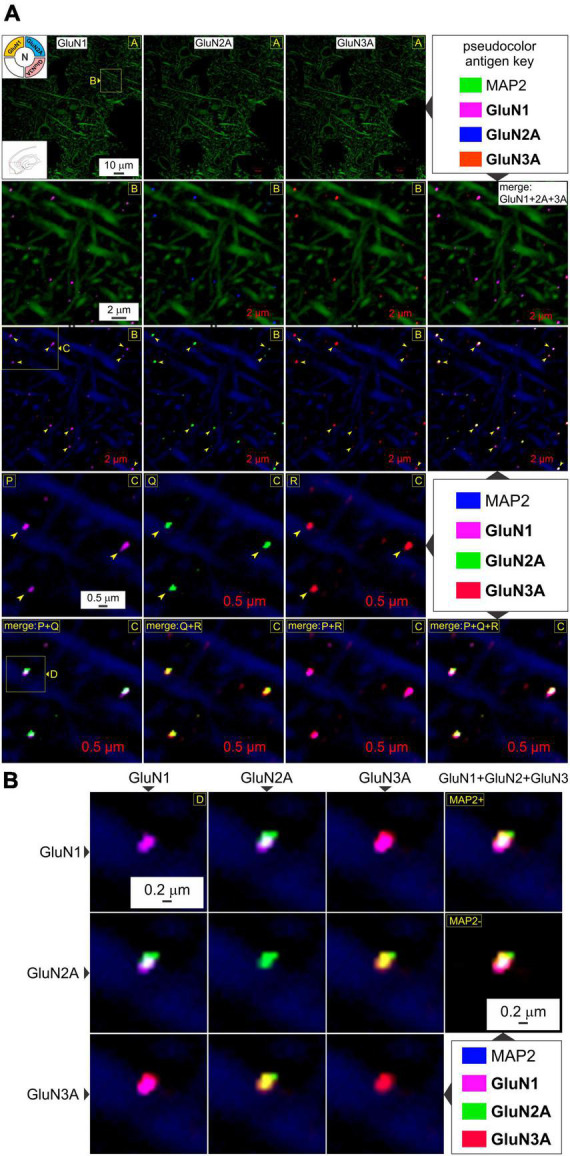
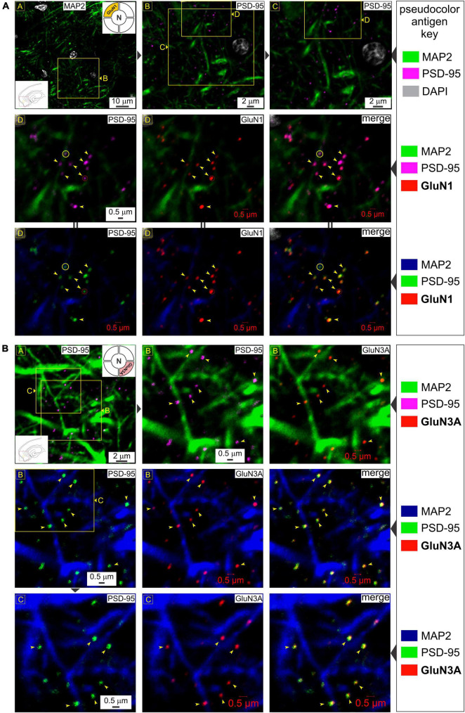
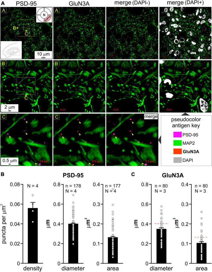
N-methyl-D-aspartate receptors (NMDARs) are one of three ligand-gated ionotropic channels that transduce the effects of neurotransmitter glutamate at excitatory synapses within the central nervous system. Their ability to influx Ca2+ into cells, unlike mature AMPA or kainate receptors, implicates them in a variety of processes ranging from synaptic plasticity to cell death. Many of the receptor's capabilities, including binding glutamate and regulating Ca2+ influx, have been attributed to their subunit composition, determined putatively using cell biology, electrophysiology and/or pharmacology. Here, we show that subunit composition of synaptic NMDARs can also be readily visualized in acute brain slices (rat) using highly specific antibodies directed against extracellular epitopes of the subunit proteins and high-resolution confocal microscopy. This has helped confirm the expression of triheteromeric t-NMDARs (containing GluN1, GluN2, and GluN3 subunits) at synapses for the first time and reconcile functional differences with diheteromeric d-NMDARs (containing GluN1 and GluN2 subunits) described previously. Even though structural information about individual receptors is still diffraction limited, fluorescently tagged receptor subunit puncta coalesce with precision at various magnifications and/or with the postsynaptic density (PSD-95) but not the presynaptic active zone marker Bassoon. These data are particularly relevant for identifying GluN3A-containing t-NMDARs that are highly Ca2+ permeable and whose expression at excitatory synapses renders neurons vulnerable to excitotoxicity and cell death. Imaging NMDAR subunit proteins at synapses not only offers firsthand insights into subunit composition to correlate function but may also help identify zones of vulnerability within brain structures underlying neurodegenerative diseases like Temporal Lobe Epilepsy.


 分享
分享
 求助内容:
求助内容: 应助结果提醒方式:
 扫码关注我们
