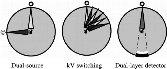
{"title":"Dual-energy CT for gastrointestinal bleeding.","authors":"Miyuki Okamura-Kawasaki, Yuya Uesugi, Satoshi Yabusaki","doi":"10.1259/bjro.20220054","DOIUrl":null,"url":null,"abstract":"<p><p>Dual-energy computed tomography (DECT) can be used for various types of analyses, including iodine quantification, and its usefulness in diagnosing gastrointestinal diseases has been reported. This pictorial review describes the use of DECT in the diagnosis of gastrointestinal bleeding. Virtual non-contrast computed tomography (CT) is available in DECT and can be used as a substitute for pre-contrast CT in the case of gastrointestinal haemorrhage. The omission of pre-contrast CT can reduce radiation exposure by approximately 30%. A low-keV virtual monochromatic X-ray image (VMI) can increase the contrast of iodine, and iodine maps can provide better visibility of extravasation. These analytical images can provide a diagnosis with a high degree of confidence. In addition, the low-keV VMI clearly illustrates the vascular structure, which may be useful for improving the visibility of vascular lesions and for confirming the arterial anatomy before embolisation. Considering that these analytical images are created on the basis of contrast-enhanced CT, the positional information of organs is entirely identical, thus allowing the comparison of images regardless of intestinal peristalsis or body motion. In conclusion, the analytical images of DECT can solve the problems of conventional protocols, and DECT is considered useful in the imaging diagnosis of gastrointestinal bleeding.</p>","PeriodicalId":72419,"journal":{"name":"BJR open","volume":"5 1","pages":"20220054"},"PeriodicalIF":2.1000,"publicationDate":"2023-01-01","publicationTypes":"Journal Article","fieldsOfStudy":null,"isOpenAccess":false,"openAccessPdf":"https://www.ncbi.nlm.nih.gov/pmc/articles/PMC10077409/pdf/","citationCount":"1","resultStr":null,"platform":"Semanticscholar","paperid":null,"PeriodicalName":"BJR open","FirstCategoryId":"1085","ListUrlMain":"https://doi.org/10.1259/bjro.20220054","RegionNum":0,"RegionCategory":null,"ArticlePicture":[],"TitleCN":null,"AbstractTextCN":null,"PMCID":null,"EPubDate":"","PubModel":"","JCR":"","JCRName":"","Score":null,"Total":0}
引用次数: 1
Abstract
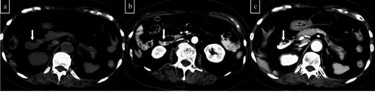
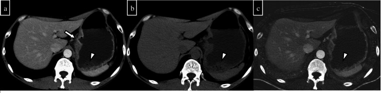
Dual-energy computed tomography (DECT) can be used for various types of analyses, including iodine quantification, and its usefulness in diagnosing gastrointestinal diseases has been reported. This pictorial review describes the use of DECT in the diagnosis of gastrointestinal bleeding. Virtual non-contrast computed tomography (CT) is available in DECT and can be used as a substitute for pre-contrast CT in the case of gastrointestinal haemorrhage. The omission of pre-contrast CT can reduce radiation exposure by approximately 30%. A low-keV virtual monochromatic X-ray image (VMI) can increase the contrast of iodine, and iodine maps can provide better visibility of extravasation. These analytical images can provide a diagnosis with a high degree of confidence. In addition, the low-keV VMI clearly illustrates the vascular structure, which may be useful for improving the visibility of vascular lesions and for confirming the arterial anatomy before embolisation. Considering that these analytical images are created on the basis of contrast-enhanced CT, the positional information of organs is entirely identical, thus allowing the comparison of images regardless of intestinal peristalsis or body motion. In conclusion, the analytical images of DECT can solve the problems of conventional protocols, and DECT is considered useful in the imaging diagnosis of gastrointestinal bleeding.


 分享
分享

 求助内容:
求助内容: 应助结果提醒方式:
 扫码关注我们
