Rakan Al-Rashdan, Mohammed Aljaberi, Ali Mohamedkhair, Akram Al-Ibraheem
{"title":"Prominent <sup>18</sup>F-FDG Uptake in the Adrenal Gland after Contralateral Adrenalectomy in a Known Case of Adrenocortical Oncocytic Carcinoma.","authors":"Rakan Al-Rashdan, Mohammed Aljaberi, Ali Mohamedkhair, Akram Al-Ibraheem","doi":"10.22038/AOJNMB.2022.67950.1472","DOIUrl":null,"url":null,"abstract":"<p><p>Adrenocortical carcinoma (ACC) is a rare type of cancer that is associated with a high rate of recurrence and poor prognosis. The main diagnostic approaches to adrenocortical cancer include CT scan, MRI and the promising role of <sup>18</sup>F-FDG PET/CT. The main therapeutic approaches include radical surgery of local disease and recurrences, as well as adjuvant mitotane therapy. The evaluation of adrenocortical carcinoma (ACC) could be difficult by using <sup>18</sup>F-FDG PET/CT in view of the significant association between the <sup>18</sup>F-FDG uptake and ACC. At the same time, not all adrenal glands with <sup>18</sup>F-FDG uptake are considered to be malignant, so awareness of these various findings is substantial for ACC management, especially with limited data regarding the role of <sup>18</sup>F-FDG PET/CT in ACC post-operative settings. This report discusses the case of a 47-year-old man with a history of left adrenocortical carcinoma who underwent adrenalectomy and received adjuvant mitotane therapy. 9 months after the surgery, a follow-up <sup>18</sup>F-FDG PET/CT scan showed that the <sup>18</sup>F-FDG uptake was prominent in the right adrenal gland without corresponding abnormal CT scan findings.</p>","PeriodicalId":72309,"journal":{"name":"","volume":"11 2","pages":"181-184"},"PeriodicalIF":0.0,"publicationDate":"2023-01-01","publicationTypes":"Journal Article","fieldsOfStudy":null,"isOpenAccess":false,"openAccessPdf":"https://www.ncbi.nlm.nih.gov/pmc/articles/PMC10261690/pdf/","citationCount":"0","resultStr":null,"platform":"Semanticscholar","paperid":null,"PeriodicalName":"","FirstCategoryId":"1085","ListUrlMain":"https://doi.org/10.22038/AOJNMB.2022.67950.1472","RegionNum":0,"RegionCategory":null,"ArticlePicture":[],"TitleCN":null,"AbstractTextCN":null,"PMCID":null,"EPubDate":"","PubModel":"","JCR":"","JCRName":"","Score":null,"Total":0}
引用次数: 0
Abstract
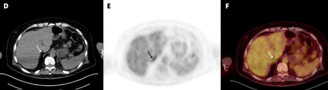
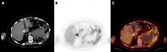
Adrenocortical carcinoma (ACC) is a rare type of cancer that is associated with a high rate of recurrence and poor prognosis. The main diagnostic approaches to adrenocortical cancer include CT scan, MRI and the promising role of 18F-FDG PET/CT. The main therapeutic approaches include radical surgery of local disease and recurrences, as well as adjuvant mitotane therapy. The evaluation of adrenocortical carcinoma (ACC) could be difficult by using 18F-FDG PET/CT in view of the significant association between the 18F-FDG uptake and ACC. At the same time, not all adrenal glands with 18F-FDG uptake are considered to be malignant, so awareness of these various findings is substantial for ACC management, especially with limited data regarding the role of 18F-FDG PET/CT in ACC post-operative settings. This report discusses the case of a 47-year-old man with a history of left adrenocortical carcinoma who underwent adrenalectomy and received adjuvant mitotane therapy. 9 months after the surgery, a follow-up 18F-FDG PET/CT scan showed that the 18F-FDG uptake was prominent in the right adrenal gland without corresponding abnormal CT scan findings.


 分享
分享
 求助内容:
求助内容: 应助结果提醒方式:
 扫码关注我们
