Comparative Study of Intraoperative Fluorescein and Indocyanine Green Videoangiography for Ruptured Cerebral Aneurysms Clipping: A Single Centre Study of 30 Cases.
{"title":"Comparative Study of Intraoperative Fluorescein and Indocyanine Green Videoangiography for Ruptured Cerebral Aneurysms Clipping: A Single Centre Study of 30 Cases.","authors":"Deepak Kumar Singh, Gaurav Sharma, Vipin Kumar Chand, Mohammad Kaif, Kuldeep Yadav","doi":"10.1055/s-0042-1751006","DOIUrl":null,"url":null,"abstract":"<p><p><b>Aim</b> This study assesses the application of microscope integrated videoangiography techniques in aneurysm clipping surgery using Indocyanine Green and Fluorescein fluorophores and evaluates merits and demerits of each technique. <b>Materials and Methods</b> Total 30 patients of cerebral aneurysmal clipping were included. Standard microsurgical procedures were done. After clipping, we administered a 25 mg bolus intravenous dose of indocyanine green with microscope focused through the INFRARED 800 camera module, followed by administration of 60 mg bolus intravenous dose of fluorescein with microscope focused through the yellow 560 module and images were assessed. <b>Results</b> The average aneurysm size was 17 mm. In 12 patients (40%), FL-VA allowed better assessment of perforating arteries (seven cases) or distal branches (three cases) or both (two cases), when compared with ICG-VA. In one case of MCA (M1) aneurysm, ICG-VA showed no fluorescent signal in one of the distal trunks whereas FL-VA showed normal signal. In one case of ACOM aneurysm, perforators were missed on ICG-VA but were seen on FL-VA. FL-VA was able to identify inadequate aneurysm clipping in one case. In two patients, FL-VA provided the advantage of real-time manipulation of the vessels to expose the vessels and aneurysms of interest. Fluorescein detected all the perforators that were visible under white light (68/68) whereas ICG was able to detect 56 (82.35%) perforators ( <i>p</i> -value< 0.05). <b>Conclusion</b> Intraoperative ICG and Fluorescein videoangiography recognize inadequate occlusion of aneurysm, decreased flow in branches or perforators. When various study parameters were considered such as ability to assess small size perforators, branching vessels, adequacy of aneurysmal clipping, and useful information on repeat imaging, FL-VA was found superior to ICG-VA.</p>","PeriodicalId":8521,"journal":{"name":"Asian Journal of Neurosurgery","volume":"18 1","pages":"25-29"},"PeriodicalIF":0.0000,"publicationDate":"2023-03-01","publicationTypes":"Journal Article","fieldsOfStudy":null,"isOpenAccess":false,"openAccessPdf":"https://ftp.ncbi.nlm.nih.gov/pub/pmc/oa_pdf/1e/8d/10-1055-s-0042-1751006.PMC10089731.pdf","citationCount":"1","resultStr":null,"platform":"Semanticscholar","paperid":null,"PeriodicalName":"Asian Journal of Neurosurgery","FirstCategoryId":"1085","ListUrlMain":"https://doi.org/10.1055/s-0042-1751006","RegionNum":0,"RegionCategory":null,"ArticlePicture":[],"TitleCN":null,"AbstractTextCN":null,"PMCID":null,"EPubDate":"","PubModel":"","JCR":"","JCRName":"","Score":null,"Total":0}
引用次数: 1
Abstract
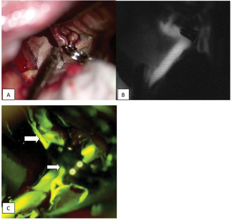
Aim This study assesses the application of microscope integrated videoangiography techniques in aneurysm clipping surgery using Indocyanine Green and Fluorescein fluorophores and evaluates merits and demerits of each technique. Materials and Methods Total 30 patients of cerebral aneurysmal clipping were included. Standard microsurgical procedures were done. After clipping, we administered a 25 mg bolus intravenous dose of indocyanine green with microscope focused through the INFRARED 800 camera module, followed by administration of 60 mg bolus intravenous dose of fluorescein with microscope focused through the yellow 560 module and images were assessed. Results The average aneurysm size was 17 mm. In 12 patients (40%), FL-VA allowed better assessment of perforating arteries (seven cases) or distal branches (three cases) or both (two cases), when compared with ICG-VA. In one case of MCA (M1) aneurysm, ICG-VA showed no fluorescent signal in one of the distal trunks whereas FL-VA showed normal signal. In one case of ACOM aneurysm, perforators were missed on ICG-VA but were seen on FL-VA. FL-VA was able to identify inadequate aneurysm clipping in one case. In two patients, FL-VA provided the advantage of real-time manipulation of the vessels to expose the vessels and aneurysms of interest. Fluorescein detected all the perforators that were visible under white light (68/68) whereas ICG was able to detect 56 (82.35%) perforators ( p -value< 0.05). Conclusion Intraoperative ICG and Fluorescein videoangiography recognize inadequate occlusion of aneurysm, decreased flow in branches or perforators. When various study parameters were considered such as ability to assess small size perforators, branching vessels, adequacy of aneurysmal clipping, and useful information on repeat imaging, FL-VA was found superior to ICG-VA.


 分享
分享

 求助内容:
求助内容: 应助结果提醒方式:
 扫码关注我们
