{"title":"Myotomy and Selective Peripheral Denervation Based on <sup>18</sup>F-FDG PET/CT in Intractable Cervical Dystonia: A Case Report.","authors":"Isamu Miura, Shiro Horisawa, Takakazu Kawamata, Takaomi Taira","doi":"10.2176/jns-nmc.2022-0392","DOIUrl":null,"url":null,"abstract":"Cervical dystonia, characterized by the involuntary contraction of cervical muscles, is the most common form of adult dystonia. In a patient with intractable cervical dystonia, we carried out a myotomy of the left obliquus capitis inferior and selective peripheral denervation (SPD) of the posterior branches of the C3-C6 spinal nerves based on preoperative 18F-fluorodeoxyglucose (18F-FDG) positron emission tomography/computed tomography (PET/CT). The patient was a 65-year-old, right-handed man with an unremarkable medical history. His head rotated involuntarily to the left. Medication and botulinum toxin injections were ineffective, and surgical treatment was considered. 18F-FDG PET/CT imaging revealed FDG uptake in the left obliquus capitis inferior, right sternocleidomastoideus, and left splenius capitis. Myotomy of the left obliquus capitis inferior and SPD of the posterior branches of the C3-C6 spinal nerves was performed under general anesthesia. During the 6-month follow-up, the patient's Toronto Western Spasmodic Torticollis Rating Scale score improved from 35 to 9. This case shows that preoperative 18F-FDG PET/CT is effective in identifying dystonic muscles and determining the surgical strategy for cervical dystonia.","PeriodicalId":19260,"journal":{"name":"NMC Case Report Journal","volume":"10 ","pages":"99-102"},"PeriodicalIF":0.0000,"publicationDate":"2023-01-01","publicationTypes":"Journal Article","fieldsOfStudy":null,"isOpenAccess":false,"openAccessPdf":"https://ftp.ncbi.nlm.nih.gov/pub/pmc/oa_pdf/5a/bb/2188-4226-10-0099.PMC10149141.pdf","citationCount":"0","resultStr":null,"platform":"Semanticscholar","paperid":null,"PeriodicalName":"NMC Case Report Journal","FirstCategoryId":"1085","ListUrlMain":"https://doi.org/10.2176/jns-nmc.2022-0392","RegionNum":0,"RegionCategory":null,"ArticlePicture":[],"TitleCN":null,"AbstractTextCN":null,"PMCID":null,"EPubDate":"","PubModel":"","JCR":"","JCRName":"","Score":null,"Total":0}
引用次数: 0
Abstract
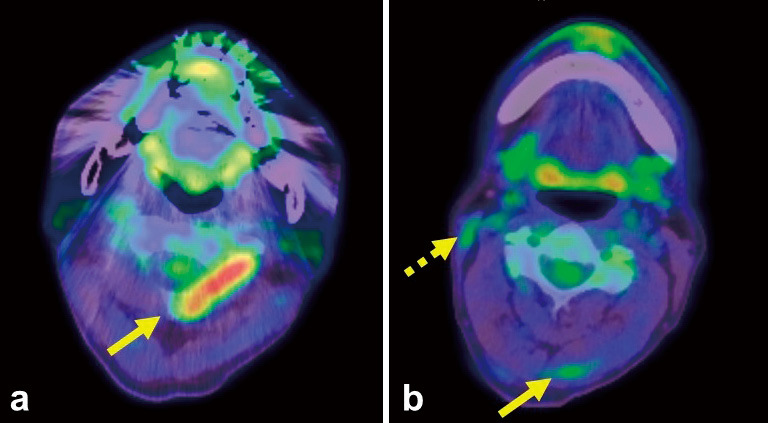
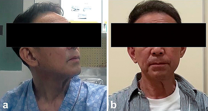
Cervical dystonia, characterized by the involuntary contraction of cervical muscles, is the most common form of adult dystonia. In a patient with intractable cervical dystonia, we carried out a myotomy of the left obliquus capitis inferior and selective peripheral denervation (SPD) of the posterior branches of the C3-C6 spinal nerves based on preoperative 18F-fluorodeoxyglucose (18F-FDG) positron emission tomography/computed tomography (PET/CT). The patient was a 65-year-old, right-handed man with an unremarkable medical history. His head rotated involuntarily to the left. Medication and botulinum toxin injections were ineffective, and surgical treatment was considered. 18F-FDG PET/CT imaging revealed FDG uptake in the left obliquus capitis inferior, right sternocleidomastoideus, and left splenius capitis. Myotomy of the left obliquus capitis inferior and SPD of the posterior branches of the C3-C6 spinal nerves was performed under general anesthesia. During the 6-month follow-up, the patient's Toronto Western Spasmodic Torticollis Rating Scale score improved from 35 to 9. This case shows that preoperative 18F-FDG PET/CT is effective in identifying dystonic muscles and determining the surgical strategy for cervical dystonia.


 分享
分享

 求助内容:
求助内容: 应助结果提醒方式:
 扫码关注我们
