Morphology variations with medial cuneiform in hallux valgus

IF 1.2 4区 医学 Q3 ANATOMY & MORPHOLOGY Anatomical Science International Pub Date : 2023-07-15 DOI:10.1007/s12565-023-00734-6
Ruining Li, Kai Tan, Yun Xie, Fasheng Wang
{"title":"Morphology variations with medial cuneiform in hallux valgus","authors":"Ruining Li,&nbsp;Kai Tan,&nbsp;Yun Xie,&nbsp;Fasheng Wang","doi":"10.1007/s12565-023-00734-6","DOIUrl":null,"url":null,"abstract":"<div><p>The hypermobility of the first tarsometatarsal joint has been identified as a key factor in the development of hallux valgus. Previous research found a link between the tarsometatarsal joint obliquity and the hallux valgus angle. Nevertheless, most studies relied on radiographs that lack 3D evidence. This study used 3D analysis to investigate the morphological differences in the medial cuneiform between hallux valgus and normal feet. In this study, twenty-three hallux valgus feet and twenty-three normal feet were scanned with computed tomography and 3D models of medial cuneiforms were reconstructed. Medial cuneonavicular and the first tarsometatarsal joint surfaces of the medial cuneiform were manually extracted. To obtain the obliquity angle of the medial cuneiform and curvature of the medial cuneonavicular joint, the joint surfaces were approximated to planes and spheres. Furthermore, the orientations of two joint surfaces were accessed through a novel positioning method. No significant difference was found in the cuneiform obliquity between hallux valgus and normal feet. Hallux valgus and normal groups did not differ significantly in any of the medial cuneiform joint orientations. The medial cuneiform in hallux valgus had a larger curvature diameter of the medial cuneonavicular joint (<i>P</i> = 0.029), indicating a flatter surface. The results demonstrated that the generally supported atavism (i.e., tarsometatarsal joint obliquity) does not exist in the hallux valgus feet. A flatter medial cuneonavicular joint surface was found in hallux valgus feet. This study contributes to the comprehensive understanding of the etiological factors with hallux valgus.</p></div>","PeriodicalId":7816,"journal":{"name":"Anatomical Science International","volume":"99 1","pages":"59 - 67"},"PeriodicalIF":1.2000,"publicationDate":"2023-07-15","publicationTypes":"Journal Article","fieldsOfStudy":null,"isOpenAccess":false,"openAccessPdf":"","citationCount":"0","resultStr":null,"platform":"Semanticscholar","paperid":null,"PeriodicalName":"Anatomical Science International","FirstCategoryId":"3","ListUrlMain":"https://link.springer.com/article/10.1007/s12565-023-00734-6","RegionNum":4,"RegionCategory":"医学","ArticlePicture":[],"TitleCN":null,"AbstractTextCN":null,"PMCID":null,"EPubDate":"","PubModel":"","JCR":"Q3","JCRName":"ANATOMY & MORPHOLOGY","Score":null,"Total":0}
引用次数: 0

Abstract

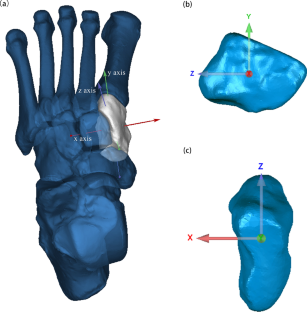
The hypermobility of the first tarsometatarsal joint has been identified as a key factor in the development of hallux valgus. Previous research found a link between the tarsometatarsal joint obliquity and the hallux valgus angle. Nevertheless, most studies relied on radiographs that lack 3D evidence. This study used 3D analysis to investigate the morphological differences in the medial cuneiform between hallux valgus and normal feet. In this study, twenty-three hallux valgus feet and twenty-three normal feet were scanned with computed tomography and 3D models of medial cuneiforms were reconstructed. Medial cuneonavicular and the first tarsometatarsal joint surfaces of the medial cuneiform were manually extracted. To obtain the obliquity angle of the medial cuneiform and curvature of the medial cuneonavicular joint, the joint surfaces were approximated to planes and spheres. Furthermore, the orientations of two joint surfaces were accessed through a novel positioning method. No significant difference was found in the cuneiform obliquity between hallux valgus and normal feet. Hallux valgus and normal groups did not differ significantly in any of the medial cuneiform joint orientations. The medial cuneiform in hallux valgus had a larger curvature diameter of the medial cuneonavicular joint (P = 0.029), indicating a flatter surface. The results demonstrated that the generally supported atavism (i.e., tarsometatarsal joint obliquity) does not exist in the hallux valgus feet. A flatter medial cuneonavicular joint surface was found in hallux valgus feet. This study contributes to the comprehensive understanding of the etiological factors with hallux valgus.
Abstract Image

查看原文
分享 分享
微信好友 朋友圈 QQ好友 复制链接
本刊更多论文
拇指外翻时内侧楔形的形态变化。
第一跖跗关节的过度活动已被确认为导致拇指外翻的关键因素。以往的研究发现,跗跖关节斜度与拇指外翻角度之间存在联系。然而,大多数研究都依赖于缺乏三维证据的X光片。本研究采用三维分析方法来研究内侧楔形骨在外翻足和正常足之间的形态差异。本研究采用计算机断层扫描技术扫描了23只拇指外翻足和23只正常足,并重建了内侧楔形的三维模型。人工提取内侧楔形关节面和第一跖跗关节面。为了获得内侧楔形关节的斜角和内侧楔形关节的曲率,将关节面近似为平面和球面。此外,还通过一种新颖的定位方法获得了两个关节面的方向。结果显示,拇指外翻组和正常组的楔形关节斜度无明显差异。拇指外翻组和正常组在任何内侧楔形关节方向上都没有明显差异。拇指外翻者的内侧楔形关节与内侧楔骨关节的曲率直径较大(P = 0.029),表明其表面较平坦。结果表明,一般认为的先天缺陷(即跖跗关节倾斜)在拇指外翻足中并不存在。研究发现,外翻足的楔形关节内侧表面较平坦。这项研究有助于全面了解足外翻的病因。
本文章由计算机程序翻译,如有差异,请以英文原文为准。
求助全文
约1分钟内获得全文 去求助
来源期刊
Anatomical Science International
Anatomical Science International 医学-解剖学与形态学
CiteScore
2.80
自引率
8.30%
发文量
50
审稿时长
>12 weeks
期刊介绍: The official English journal of the Japanese Association of Anatomists, Anatomical Science International (formerly titled Kaibogaku Zasshi) publishes original research articles dealing with morphological sciences. Coverage in the journal includes molecular, cellular, histological and gross anatomical studies on humans and on normal and experimental animals, as well as functional morphological, biochemical, physiological and behavioral studies if they include morphological analysis.
期刊最新文献
Prevalence and clinical aspects of os trigonum: a meta-analysis. Variant anatomy of the iliac veins and presence of two venous rings around the arteries in the pelvis. Regulatory mechanisms governing GLI proteins in hedgehog signaling. Envisioning the future of ASI: reflections on the 100th anniversary. Unilateral two-headed extensor digitorum longus muscle: atypical configuration and clinical implications.
×
引用
GB/T 7714-2015
复制
MLA
复制
APA
复制
导出至
BibTeX EndNote RefMan NoteFirst NoteExpress
×
×
提示
您的信息不完整,为了账户安全,请先补充。
现在去补充
×
提示
您因"违规操作"
具体请查看互助需知
我知道了
×
提示
现在去查看 取消
×
提示
确定
0
微信
客服QQ
Book学术公众号 扫码关注我们
反馈
×
意见反馈
请填写您的意见或建议
请填写您的手机或邮箱
已复制链接
已复制链接
快去分享给好友吧!
我知道了
×
扫码分享
扫码分享
Book学术官方微信
Book学术文献互助
Book学术文献互助群
群 号:481959085
Book学术
文献互助 智能选刊 最新文献 互助须知 联系我们:info@booksci.cn
Book学术提供免费学术资源搜索服务,方便国内外学者检索中英文文献。致力于提供最便捷和优质的服务体验。
Copyright © 2023 Book学术 All rights reserved.
ghs 京公网安备 11010802042870号 京ICP备2023020795号-1