Miguel Mascarenhas, Rui Morais, Regina Teixeira, Guilherme Macedo
{"title":"A case report of an unexpected colonic polyp: cavernous hemangioma.","authors":"Miguel Mascarenhas, Rui Morais, Regina Teixeira, Guilherme Macedo","doi":"10.3393/ac.2020.00535.0076","DOIUrl":null,"url":null,"abstract":"<p><p>Cavernous hemangiomas of the colon are rare, benign vascular lesions, and the site most commonly affected is the rectosigmoid junction. Surgical treatment is recommended for large diffuse lesions but in the presence of pedunculated lesions, endoscopic resection should be preferred if possible. We report a case of a 65-year-old man referred for colonoscopy after positive fecal occult blood, that revealed at the level of the sigmoid colon, a wide base pedunculated polyp (35 mm) occupying more than half of the lumen, with the covering mucosa with a vinous appearance. In order to remove the lesion, a detachable snare was placed and polypectomy was performed. During the procedure, the detachable snare was cut with active bleeding, controlled after clip placement and diluted adrenaline injection. Afterwards, histology revealed a polypoid lesion with a hyperplastic mucosa and submucosal plane expanded by numerous thick-walled vessels in the context of a cavernous colonic hemangioma.</p>","PeriodicalId":8267,"journal":{"name":"Annals of Coloproctology","volume":"39 3","pages":"280-282"},"PeriodicalIF":2.1000,"publicationDate":"2023-06-01","publicationTypes":"Journal Article","fieldsOfStudy":null,"isOpenAccess":false,"openAccessPdf":"https://ftp.ncbi.nlm.nih.gov/pub/pmc/oa_pdf/8b/b7/ac-2020-00535-0076.PMC10338163.pdf","citationCount":"1","resultStr":null,"platform":"Semanticscholar","paperid":null,"PeriodicalName":"Annals of Coloproctology","FirstCategoryId":"1085","ListUrlMain":"https://doi.org/10.3393/ac.2020.00535.0076","RegionNum":0,"RegionCategory":null,"ArticlePicture":[],"TitleCN":null,"AbstractTextCN":null,"PMCID":null,"EPubDate":"","PubModel":"","JCR":"Q2","JCRName":"GASTROENTEROLOGY & HEPATOLOGY","Score":null,"Total":0}
引用次数: 1
Abstract
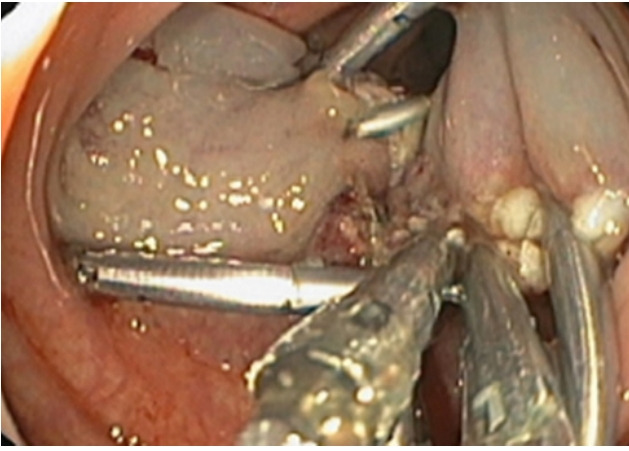
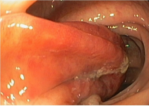
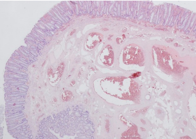
Cavernous hemangiomas of the colon are rare, benign vascular lesions, and the site most commonly affected is the rectosigmoid junction. Surgical treatment is recommended for large diffuse lesions but in the presence of pedunculated lesions, endoscopic resection should be preferred if possible. We report a case of a 65-year-old man referred for colonoscopy after positive fecal occult blood, that revealed at the level of the sigmoid colon, a wide base pedunculated polyp (35 mm) occupying more than half of the lumen, with the covering mucosa with a vinous appearance. In order to remove the lesion, a detachable snare was placed and polypectomy was performed. During the procedure, the detachable snare was cut with active bleeding, controlled after clip placement and diluted adrenaline injection. Afterwards, histology revealed a polypoid lesion with a hyperplastic mucosa and submucosal plane expanded by numerous thick-walled vessels in the context of a cavernous colonic hemangioma.


 分享
分享
 求助内容:
求助内容: 应助结果提醒方式:
 扫码关注我们
