{"title":"MMP-3-mediated cleavage of OPN is involved in copper oxide nanoparticle-induced activation of fibroblasts.","authors":"Yuanbao Zhang, Yiqun Mo, Yue Zhang, Jiali Yuan, Qunwei Zhang","doi":"10.1186/s12989-023-00532-y","DOIUrl":null,"url":null,"abstract":"<p><strong>Background: </strong>Copper oxide nanoparticles (Nano-CuO) are one of the most produced and used nanomaterials. Previous studies have shown that exposure to Nano-CuO caused acute lung injury, inflammation, and fibrosis. However, the mechanisms underlying Nano-CuO-induced lung fibrosis are still unclear. Here, we hypothesized that exposure of human lung epithelial cells and macrophages to Nano-CuO would upregulate MMP-3, which cleaved osteopontin (OPN), resulting in fibroblast activation and lung fibrosis.</p><p><strong>Methods: </strong>A triple co-culture model was established to explore the mechanisms underlying Nano-CuO-induced fibroblast activation. Cytotoxicity of Nano-CuO on BEAS-2B, U937* macrophages, and MRC-5 fibroblasts were determined by alamarBlue and MTS assays. The expression or activity of MMP-3, OPN, and fibrosis-associated proteins was determined by Western blot or zymography assay. Migration of MRC-5 fibroblasts was evaluated by wound healing assay. MMP-3 siRNA and an RGD-containing peptide, GRGDSP, were used to explore the role of MMP-3 and cleaved OPN in fibroblast activation.</p><p><strong>Results: </strong>Exposure to non-cytotoxic doses of Nano-CuO (0.5 and 1 µg/mL) caused increased expression and activity of MMP-3 in the conditioned media of BEAS-2B and U937* cells, but not MRC-5 fibroblasts. Nano-CuO exposure also caused increased production of cleaved OPN fragments, which was abolished by MMP-3 siRNA transfection. Conditioned media from Nano-CuO-exposed BEAS-2B, U937*, or the co-culture of BEAS-2B and U937* caused activation of unexposed MRC-5 fibroblasts. However, direct exposure of MRC-5 fibroblasts to Nano-CuO did not induce their activation. In a triple co-culture system, exposure of BEAS-2B and U937* cells to Nano-CuO caused activation of unexposed MRC-5 fibroblasts, while transfection of MMP-3 siRNA in BEAS-2B and U937* cells significantly inhibited the activation and migration of MRC-5 fibroblasts. In addition, pretreatment with GRGDSP peptide inhibited Nano-CuO-induced activation and migration of MRC-5 fibroblasts in the triple co-culture system.</p><p><strong>Conclusions: </strong>Our results demonstrated that Nano-CuO exposure caused increased production of MMP-3 from lung epithelial BEAS-2B cells and U937* macrophages, which cleaved OPN, resulting in the activation of lung fibroblasts MRC-5. These results suggest that MMP-3-cleaved OPN may play a key role in Nano-CuO-induced activation of lung fibroblasts. More investigations are needed to confirm whether these effects are due to the nanoparticles themselves and/or Cu ions.</p>","PeriodicalId":19847,"journal":{"name":"Particle and Fibre Toxicology","volume":"20 1","pages":"22"},"PeriodicalIF":8.2000,"publicationDate":"2023-05-22","publicationTypes":"Journal Article","fieldsOfStudy":null,"isOpenAccess":false,"openAccessPdf":"https://www.ncbi.nlm.nih.gov/pmc/articles/PMC10201731/pdf/","citationCount":"0","resultStr":null,"platform":"Semanticscholar","paperid":null,"PeriodicalName":"Particle and Fibre Toxicology","FirstCategoryId":"3","ListUrlMain":"https://doi.org/10.1186/s12989-023-00532-y","RegionNum":1,"RegionCategory":"医学","ArticlePicture":[],"TitleCN":null,"AbstractTextCN":null,"PMCID":null,"EPubDate":"","PubModel":"","JCR":"Q1","JCRName":"TOXICOLOGY","Score":null,"Total":0}
引用次数: 0
Abstract
Background: Copper oxide nanoparticles (Nano-CuO) are one of the most produced and used nanomaterials. Previous studies have shown that exposure to Nano-CuO caused acute lung injury, inflammation, and fibrosis. However, the mechanisms underlying Nano-CuO-induced lung fibrosis are still unclear. Here, we hypothesized that exposure of human lung epithelial cells and macrophages to Nano-CuO would upregulate MMP-3, which cleaved osteopontin (OPN), resulting in fibroblast activation and lung fibrosis.
Methods: A triple co-culture model was established to explore the mechanisms underlying Nano-CuO-induced fibroblast activation. Cytotoxicity of Nano-CuO on BEAS-2B, U937* macrophages, and MRC-5 fibroblasts were determined by alamarBlue and MTS assays. The expression or activity of MMP-3, OPN, and fibrosis-associated proteins was determined by Western blot or zymography assay. Migration of MRC-5 fibroblasts was evaluated by wound healing assay. MMP-3 siRNA and an RGD-containing peptide, GRGDSP, were used to explore the role of MMP-3 and cleaved OPN in fibroblast activation.
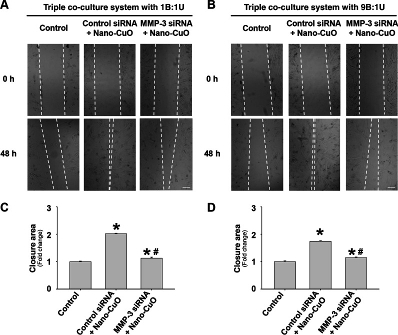
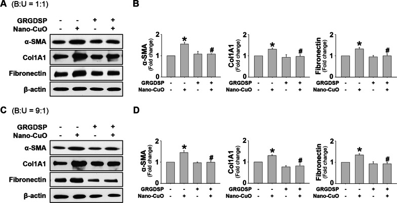
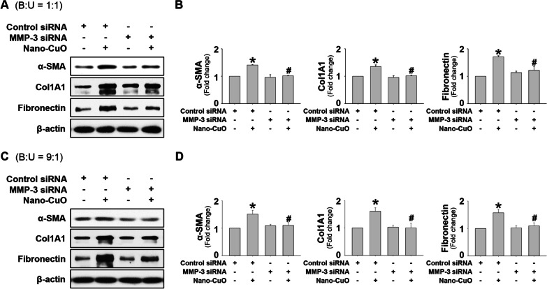
Results: Exposure to non-cytotoxic doses of Nano-CuO (0.5 and 1 µg/mL) caused increased expression and activity of MMP-3 in the conditioned media of BEAS-2B and U937* cells, but not MRC-5 fibroblasts. Nano-CuO exposure also caused increased production of cleaved OPN fragments, which was abolished by MMP-3 siRNA transfection. Conditioned media from Nano-CuO-exposed BEAS-2B, U937*, or the co-culture of BEAS-2B and U937* caused activation of unexposed MRC-5 fibroblasts. However, direct exposure of MRC-5 fibroblasts to Nano-CuO did not induce their activation. In a triple co-culture system, exposure of BEAS-2B and U937* cells to Nano-CuO caused activation of unexposed MRC-5 fibroblasts, while transfection of MMP-3 siRNA in BEAS-2B and U937* cells significantly inhibited the activation and migration of MRC-5 fibroblasts. In addition, pretreatment with GRGDSP peptide inhibited Nano-CuO-induced activation and migration of MRC-5 fibroblasts in the triple co-culture system.
Conclusions: Our results demonstrated that Nano-CuO exposure caused increased production of MMP-3 from lung epithelial BEAS-2B cells and U937* macrophages, which cleaved OPN, resulting in the activation of lung fibroblasts MRC-5. These results suggest that MMP-3-cleaved OPN may play a key role in Nano-CuO-induced activation of lung fibroblasts. More investigations are needed to confirm whether these effects are due to the nanoparticles themselves and/or Cu ions.
期刊介绍:
Particle and Fibre Toxicology is an online journal that is open access and peer-reviewed. It covers a range of disciplines such as material science, biomaterials, and nanomedicine, focusing on the toxicological effects of particles and fibres. The journal serves as a platform for scientific debate and communication among toxicologists and scientists from different fields who work with particle and fibre materials. The main objective of the journal is to deepen our understanding of the physico-chemical properties of particles, their potential for human exposure, and the resulting biological effects. It also addresses regulatory issues related to particle exposure in workplaces and the general environment. Moreover, the journal recognizes that there are various situations where particles can pose a toxicological threat, such as the use of old materials in new applications or the introduction of new materials altogether. By encompassing all these disciplines, Particle and Fibre Toxicology provides a comprehensive source for research in this field.


 分享
分享
 求助内容:
求助内容: 应助结果提醒方式:
 扫码关注我们
