{"title":"Rare Pleomorphic Liposarcoma Presented as Jejunal Obstruction.","authors":"Mohammad Al-Attar, Anup Jnawali, Michelle Yang","doi":"10.1155/2023/8040232","DOIUrl":null,"url":null,"abstract":"<p><p>Pleomorphic liposarcoma (PLS) is typically found in the lower and upper extremities. PLS arising in the gastrointestinal (GI) tract is extremely rare. Here, we reported a case of a 71-year-old female with a history of rectal adenocarcinoma presenting with small bowel obstruction. Small bowel resection was performed and revealed a 7.8 cm transmural mass in the jejunum. Histology reviewed a heterogenous epithelioid malignant tumor with intracytoplasmic fatty droplets scalloping the nucleus consistent with lipoblasts in some cells and others with numerous PAS/diastase+intracytoplasmic eosinophilic globules. Scattered multinucleated giant cells were also present. Mitotic count was up to 80/10 HPFs including some bizarre mitotic figures, and Ki67 proliferation index was approximately 60%. Immunohistochemistry demonstrated that the malignant cells were negative for pancytokeratin, CD117, DOG1, SMA, desmin, MyoD1, ERG1, CD34, CD31, SOX10, Melan A, and S100. INI1 was retained. Beta-catenin showed normal membranous staining. P53 was diffusely positive suggestive of mutant phenotype. Fluorescence in situ hybridization (FISH) assay was negative for MDM2 amplification and DDIT3 rearrangement. The overall morphologic and immunohistochemical features supported a diagnosis of high-grade pleomorphic liposarcoma. Diagnosis of PLS can be challenging due to its rarity in GI tract and lack of specific biomarkers, and histomorphology with identification of lipoblasts remains the gold standard.</p>","PeriodicalId":45638,"journal":{"name":"Case Reports in Pathology","volume":"2023 ","pages":"8040232"},"PeriodicalIF":0.5000,"publicationDate":"2023-01-01","publicationTypes":"Journal Article","fieldsOfStudy":null,"isOpenAccess":false,"openAccessPdf":"https://www.ncbi.nlm.nih.gov/pmc/articles/PMC10319459/pdf/","citationCount":"0","resultStr":null,"platform":"Semanticscholar","paperid":null,"PeriodicalName":"Case Reports in Pathology","FirstCategoryId":"1085","ListUrlMain":"https://doi.org/10.1155/2023/8040232","RegionNum":0,"RegionCategory":null,"ArticlePicture":[],"TitleCN":null,"AbstractTextCN":null,"PMCID":null,"EPubDate":"","PubModel":"","JCR":"Q4","JCRName":"PATHOLOGY","Score":null,"Total":0}
引用次数: 0
Abstract
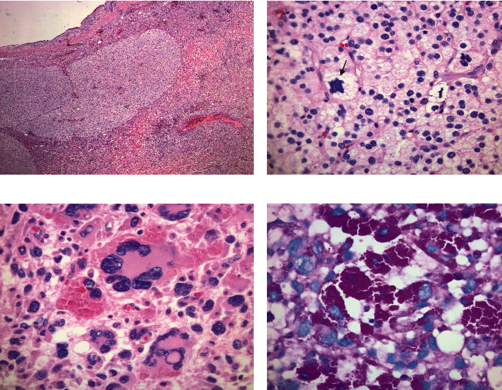
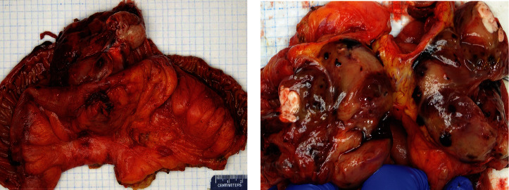
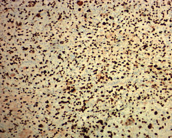
Pleomorphic liposarcoma (PLS) is typically found in the lower and upper extremities. PLS arising in the gastrointestinal (GI) tract is extremely rare. Here, we reported a case of a 71-year-old female with a history of rectal adenocarcinoma presenting with small bowel obstruction. Small bowel resection was performed and revealed a 7.8 cm transmural mass in the jejunum. Histology reviewed a heterogenous epithelioid malignant tumor with intracytoplasmic fatty droplets scalloping the nucleus consistent with lipoblasts in some cells and others with numerous PAS/diastase+intracytoplasmic eosinophilic globules. Scattered multinucleated giant cells were also present. Mitotic count was up to 80/10 HPFs including some bizarre mitotic figures, and Ki67 proliferation index was approximately 60%. Immunohistochemistry demonstrated that the malignant cells were negative for pancytokeratin, CD117, DOG1, SMA, desmin, MyoD1, ERG1, CD34, CD31, SOX10, Melan A, and S100. INI1 was retained. Beta-catenin showed normal membranous staining. P53 was diffusely positive suggestive of mutant phenotype. Fluorescence in situ hybridization (FISH) assay was negative for MDM2 amplification and DDIT3 rearrangement. The overall morphologic and immunohistochemical features supported a diagnosis of high-grade pleomorphic liposarcoma. Diagnosis of PLS can be challenging due to its rarity in GI tract and lack of specific biomarkers, and histomorphology with identification of lipoblasts remains the gold standard.


 分享
分享
 求助内容:
求助内容: 应助结果提醒方式:
 扫码关注我们
