{"title":"Distribution of the Retinal Microcirculation Based on the Morphology of Peripapillary Atrophy in High Myopia.","authors":"Wenquan Tang, Junyi Ouyang, YuLin Luo","doi":"10.1159/000531509","DOIUrl":null,"url":null,"abstract":"<p><strong>Introduction: </strong>The objective of this study was to evaluate the retinal microvasculature of the optic nerve head and macula in high myopia (HM), investigate the association between the vascular parameters and peripapillary atrophy (PPA) deformation, and assess and identify the PPA morphology changes during the development of HM.</p><p><strong>Methods: </strong>One hundred sixty-seven right eyes from 167 HM patients were enrolled in this cross-sectional study. Using the optical coherence tomography angiography (OCTA) and fundus camera, we evaluated the following parameters: radian and type of PPA, intrapapillary vascular density (IVD), peripapillary vascular density (PVD), macular vascular density (MVD), and foveal avascular zone (FAZ). Based on the PPA radian, subjects were divided into four groups: the non-PPA, temporal PPA, advanced PPA, and annular PPA. At the same time, the above parameters were compared between the groups using analysis of variance (ANOVA) and least significant difference test.</p><p><strong>Results: </strong>Total enrolled patients were divided into the non-PPA group (22 eyes), temporal-PPA group (70 eyes), advanced-PPA group (60 eyes), and annular-PPA group (15 eyes). The results showed that the PVD in the annular-PPA group was smaller than that in the non-PPA group, especially in the superonasal, nasosuperior, nasoinferior, inferotemporal, temporoinferior, and superotemporal directions (F = 4.059, 5.014, 2.830, 4.798, 5.892, 3.439; p < 0.05). Notably, the PVD showcased the highest value in temporal, followed by that in superior and inferior, and the lowest in the nasal. Concerning the fovea deep macular vascular density, FAZ area, and subfoveal choroidal thickness in the annular-PPA group, they were less than those of the rest of the groups (p < 0.05).</p><p><strong>Conclusion: </strong>The retinal microvasculature differed significantly in HM according to the PPA morphology. In addition to PVD and SFCT, the PPA can also affect FAZ. Finally, we speculated that PVD demonstrated better predictability of myopic progression than MVD.</p>","PeriodicalId":19662,"journal":{"name":"Ophthalmic Research","volume":" ","pages":"1085-1095"},"PeriodicalIF":1.9000,"publicationDate":"2023-01-01","publicationTypes":"Journal Article","fieldsOfStudy":null,"isOpenAccess":false,"openAccessPdf":"https://www.ncbi.nlm.nih.gov/pmc/articles/PMC10614452/pdf/","citationCount":"0","resultStr":null,"platform":"Semanticscholar","paperid":null,"PeriodicalName":"Ophthalmic Research","FirstCategoryId":"3","ListUrlMain":"https://doi.org/10.1159/000531509","RegionNum":4,"RegionCategory":"医学","ArticlePicture":[],"TitleCN":null,"AbstractTextCN":null,"PMCID":null,"EPubDate":"2023/7/17 0:00:00","PubModel":"Epub","JCR":"Q2","JCRName":"OPHTHALMOLOGY","Score":null,"Total":0}
引用次数: 0
Abstract
Introduction: The objective of this study was to evaluate the retinal microvasculature of the optic nerve head and macula in high myopia (HM), investigate the association between the vascular parameters and peripapillary atrophy (PPA) deformation, and assess and identify the PPA morphology changes during the development of HM.
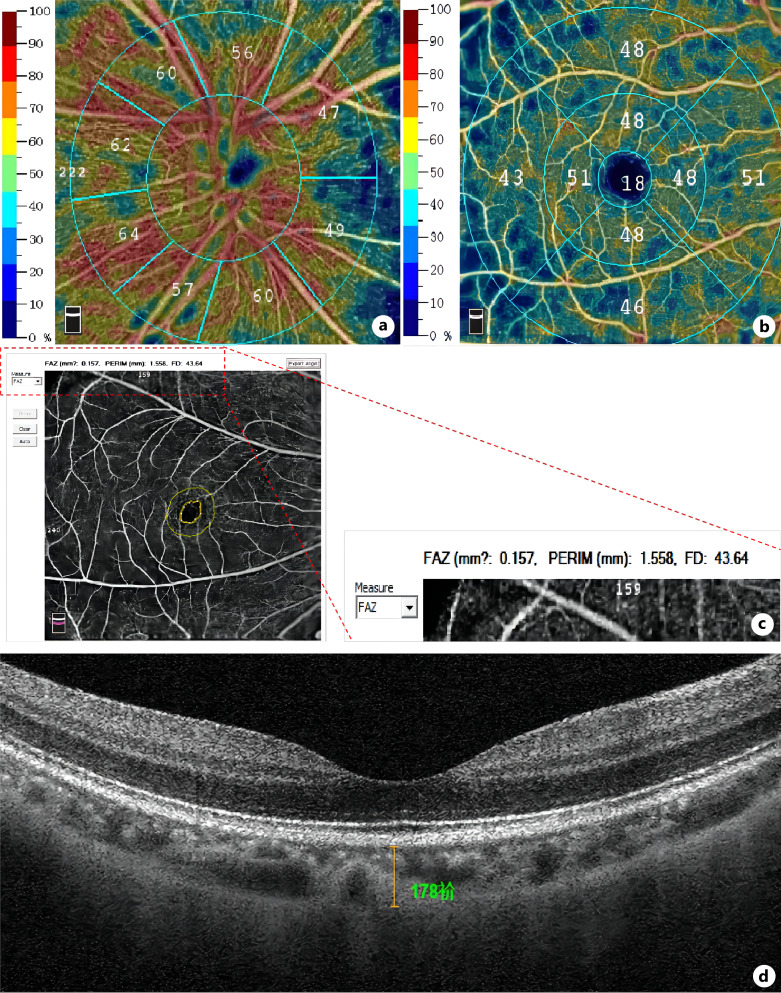
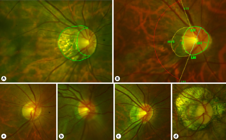
Methods: One hundred sixty-seven right eyes from 167 HM patients were enrolled in this cross-sectional study. Using the optical coherence tomography angiography (OCTA) and fundus camera, we evaluated the following parameters: radian and type of PPA, intrapapillary vascular density (IVD), peripapillary vascular density (PVD), macular vascular density (MVD), and foveal avascular zone (FAZ). Based on the PPA radian, subjects were divided into four groups: the non-PPA, temporal PPA, advanced PPA, and annular PPA. At the same time, the above parameters were compared between the groups using analysis of variance (ANOVA) and least significant difference test.
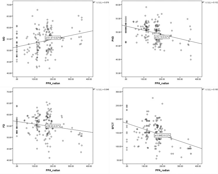
Results: Total enrolled patients were divided into the non-PPA group (22 eyes), temporal-PPA group (70 eyes), advanced-PPA group (60 eyes), and annular-PPA group (15 eyes). The results showed that the PVD in the annular-PPA group was smaller than that in the non-PPA group, especially in the superonasal, nasosuperior, nasoinferior, inferotemporal, temporoinferior, and superotemporal directions (F = 4.059, 5.014, 2.830, 4.798, 5.892, 3.439; p < 0.05). Notably, the PVD showcased the highest value in temporal, followed by that in superior and inferior, and the lowest in the nasal. Concerning the fovea deep macular vascular density, FAZ area, and subfoveal choroidal thickness in the annular-PPA group, they were less than those of the rest of the groups (p < 0.05).
Conclusion: The retinal microvasculature differed significantly in HM according to the PPA morphology. In addition to PVD and SFCT, the PPA can also affect FAZ. Finally, we speculated that PVD demonstrated better predictability of myopic progression than MVD.
期刊介绍:
''Ophthalmic Research'' features original papers and reviews reporting on translational and clinical studies. Authors from throughout the world cover research topics on every field in connection with physical, physiologic, pharmacological, biochemical and molecular biological aspects of ophthalmology. This journal also aims to provide a record of international clinical research for both researchers and clinicians in ophthalmology. Finally, the transfer of information from fundamental research to clinical research and clinical practice is particularly welcome.


 分享
分享
 求助内容:
求助内容: 应助结果提醒方式:
 扫码关注我们
