{"title":"Myopia control utilizing low-dose atropine as an isolated therapy or in combination with other optical measures: A retrospective cohort study.","authors":"Nir Erdinest, Naomi London, Itay Lavy, Nadav Levinger, Eran Pras, Yair Morad","doi":"10.4103/tjo.tjo_31_22","DOIUrl":null,"url":null,"abstract":"<p><strong>Purpose: </strong>To assess the additive potency of low-dose atropine combined with optical measures designed to decrease myopia progression.</p><p><strong>Materials and methods: </strong>This retrospective study included 104 myopic children aged 5-12 over 4 years, divided into five groups: daily instillation of 0.01% atropine and distance single-vision spectacles (A), 0.01% atropine and progressive addition lenses (A + PAL), 0.01% atropine and soft contact lens with peripheral blur (A + CL). Two control groups were included, prescribed bifocal spectacles or single vision (SV) spectacles. Cycloplegic spherical equivalence refraction was measured biannually, including 1 year after cessation of treatment.</p><p><strong>Results: </strong>A significant decrease in myopia progression was noted during the 2<sup>nd</sup> and 3<sup>rd</sup> years of atropine treatment: A -0.55 ± 0.55D, -0.15 ± 0.15, -0.12 ± 0.12D were 1<sup>st</sup>, 2<sup>nd</sup>, 3<sup>rd</sup> years, respectively, A + PAL -0.47 ± 0.37D, -0.10 ± 0.25D, and -0.11 ± 0.25D were 1<sup>st</sup>, 2<sup>nd</sup>, 3<sup>rd</sup> years, respectively, A + CL -0.36 ± 0.43D, -0.13 ± 0.29D, and -0.10 ± 0.27D were 1<sup>st</sup>, 2<sup>nd</sup>, 3<sup>rd</sup> years, respectively. Myopia progression over 3 years, respectively, was -0.82 ± 0.50D, -0.70 ± 0.69D, -0.59 ± 0.66D in the bifocal group and -1.20 ± 1.28D, -0.72 ± 0.62D, -0.65 ± 0.47D in the SV group. One year after cessation of atropine treatment, myopia progression was - 0.32 ± 0.31D in A, -0.23 ± 0.28D in A + PAL, and -0.18 ± 0.35D in A + CL.</p><p><strong>Conclusion: </strong>Atropine 0.01% presented as effective at decelerating myopia progression, more prominent in the 2<sup>nd</sup> and 3<sup>rd</sup> years of treatment. Combining atropine 0.01% with optical modalities exhibited a trend for added efficacy over monotherapy. A + CL exhibited the least rebound effect 1 year after cessation of treatment.</p>","PeriodicalId":44978,"journal":{"name":"Taiwan Journal of Ophthalmology","volume":"13 2","pages":"231-237"},"PeriodicalIF":1.2000,"publicationDate":"2023-04-01","publicationTypes":"Journal Article","fieldsOfStudy":null,"isOpenAccess":false,"openAccessPdf":"https://ftp.ncbi.nlm.nih.gov/pub/pmc/oa_pdf/de/51/TJO-13-231.PMC10361442.pdf","citationCount":"0","resultStr":null,"platform":"Semanticscholar","paperid":null,"PeriodicalName":"Taiwan Journal of Ophthalmology","FirstCategoryId":"1085","ListUrlMain":"https://doi.org/10.4103/tjo.tjo_31_22","RegionNum":0,"RegionCategory":null,"ArticlePicture":[],"TitleCN":null,"AbstractTextCN":null,"PMCID":null,"EPubDate":"","PubModel":"","JCR":"Q4","JCRName":"OPHTHALMOLOGY","Score":null,"Total":0}
引用次数: 0
Abstract
Purpose: To assess the additive potency of low-dose atropine combined with optical measures designed to decrease myopia progression.
Materials and methods: This retrospective study included 104 myopic children aged 5-12 over 4 years, divided into five groups: daily instillation of 0.01% atropine and distance single-vision spectacles (A), 0.01% atropine and progressive addition lenses (A + PAL), 0.01% atropine and soft contact lens with peripheral blur (A + CL). Two control groups were included, prescribed bifocal spectacles or single vision (SV) spectacles. Cycloplegic spherical equivalence refraction was measured biannually, including 1 year after cessation of treatment.
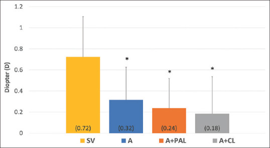
Results: A significant decrease in myopia progression was noted during the 2nd and 3rd years of atropine treatment: A -0.55 ± 0.55D, -0.15 ± 0.15, -0.12 ± 0.12D were 1st, 2nd, 3rd years, respectively, A + PAL -0.47 ± 0.37D, -0.10 ± 0.25D, and -0.11 ± 0.25D were 1st, 2nd, 3rd years, respectively, A + CL -0.36 ± 0.43D, -0.13 ± 0.29D, and -0.10 ± 0.27D were 1st, 2nd, 3rd years, respectively. Myopia progression over 3 years, respectively, was -0.82 ± 0.50D, -0.70 ± 0.69D, -0.59 ± 0.66D in the bifocal group and -1.20 ± 1.28D, -0.72 ± 0.62D, -0.65 ± 0.47D in the SV group. One year after cessation of atropine treatment, myopia progression was - 0.32 ± 0.31D in A, -0.23 ± 0.28D in A + PAL, and -0.18 ± 0.35D in A + CL.
Conclusion: Atropine 0.01% presented as effective at decelerating myopia progression, more prominent in the 2nd and 3rd years of treatment. Combining atropine 0.01% with optical modalities exhibited a trend for added efficacy over monotherapy. A + CL exhibited the least rebound effect 1 year after cessation of treatment.


 分享
分享
 求助内容:
求助内容: 应助结果提醒方式:
 扫码关注我们
