Giovanni Paolino, Matteo Riccardo Di Nicola, Riccardo Pampena, Vittoria Giulia Bianchi, Santo Raffaele Mercuri
{"title":"Reflectance Confocal Microscopy of Skin after the Sting of the Jellyfish <i>Pelagia noctiluca</i>.","authors":"Giovanni Paolino, Matteo Riccardo Di Nicola, Riccardo Pampena, Vittoria Giulia Bianchi, Santo Raffaele Mercuri","doi":"10.1159/000529049","DOIUrl":null,"url":null,"abstract":"<p><p>Jellyfish are aquatic animals of the phylum Cnidaria found in seas all over the world. They are characterized by the presence of cnidocytes, cells that contain a secretory organelle, the cnidocyst, mainly used for predation and defense purposes. An adult female patient presented to our Unit of Dermatology, for a 10 days-old history of macular-erythematous lesions in her right upper limb, due to a sting by a mauve stinger <i>Pelagia noctiluca</i>. Dermoscopy showed a general pinkish background surmounted by numerous brown dots and lines, distributed along the surface of the skin. Reflectance confocal microscopy (RCM) showed the presence of multiple partially hyperreflective, highly coiled, hollow, and harpoonlike structures through the epidermis but without the barbed tubes found in a previous RCM report, likely due to a greater time elapsed between the sting and the dermatological visit. This case highlights how dermoscopy and RCM may help clinicians for the diagnosis of jellyfish stings.</p>","PeriodicalId":9619,"journal":{"name":"Case Reports in Dermatology","volume":"15 1","pages":"105-109"},"PeriodicalIF":0.8000,"publicationDate":"2023-01-01","publicationTypes":"Journal Article","fieldsOfStudy":null,"isOpenAccess":false,"openAccessPdf":"https://ftp.ncbi.nlm.nih.gov/pub/pmc/oa_pdf/77/4d/cde-2023-0015-0001-529049.PMC10368086.pdf","citationCount":"1","resultStr":null,"platform":"Semanticscholar","paperid":null,"PeriodicalName":"Case Reports in Dermatology","FirstCategoryId":"1085","ListUrlMain":"https://doi.org/10.1159/000529049","RegionNum":0,"RegionCategory":null,"ArticlePicture":[],"TitleCN":null,"AbstractTextCN":null,"PMCID":null,"EPubDate":"","PubModel":"","JCR":"Q4","JCRName":"DERMATOLOGY","Score":null,"Total":0}
引用次数: 1
Abstract
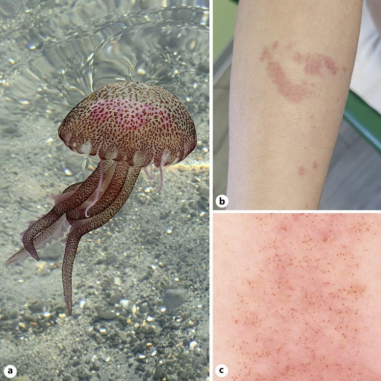
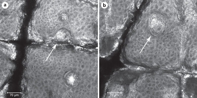
Jellyfish are aquatic animals of the phylum Cnidaria found in seas all over the world. They are characterized by the presence of cnidocytes, cells that contain a secretory organelle, the cnidocyst, mainly used for predation and defense purposes. An adult female patient presented to our Unit of Dermatology, for a 10 days-old history of macular-erythematous lesions in her right upper limb, due to a sting by a mauve stinger Pelagia noctiluca. Dermoscopy showed a general pinkish background surmounted by numerous brown dots and lines, distributed along the surface of the skin. Reflectance confocal microscopy (RCM) showed the presence of multiple partially hyperreflective, highly coiled, hollow, and harpoonlike structures through the epidermis but without the barbed tubes found in a previous RCM report, likely due to a greater time elapsed between the sting and the dermatological visit. This case highlights how dermoscopy and RCM may help clinicians for the diagnosis of jellyfish stings.


 分享
分享
 求助内容:
求助内容: 应助结果提醒方式:
 扫码关注我们
