{"title":"Examination of postmortem changes in the lungs, trachea, and bronchi in a rat model imaged with small-animal computed tomography.","authors":"Takahiro Matsuyama, Seiichiro Ota, Yoshitaka Inui, Naoko Fujii, Tetsuya Tsukamoto, Ichiro Isobe, Katsumi Tsujioka, Shizuko Nagao, Ryosuke Tanabe, Hiroshi Toyama","doi":"10.20407/fmj.2022-002","DOIUrl":null,"url":null,"abstract":"<p><strong>Objectives: </strong>As less autopsies are performed, the need for postmortem computed tomography (PMCT) as an alternative is increasing. It is important to know how postmortem changes over time are reflected on CT, in order to improve the diagnostic capability of PMCT and replace forensic pathology evaluations such as time of death estimation.</p><p><strong>Methods: </strong>In this study, we examined temporal changes on postmortem chest CT images of a rat model. After acquiring antemortem images under isoflurane inhalation anesthesia, the rats were euthanized with a rapid intravenous injection of anesthetics. From immediately after death to 48 hours postmortem, chest images were acquired using small-animal CT. The 3D images were then evaluated on a workstation to measure the antemortem and postmortem air content in the lungs, trachea, and bronchi over time.</p><p><strong>Results: </strong>The air content in the lungs decreased, but the air content of the trachea and bronchi temporarily increased 1-12 hours postmortem, then decreased at 48 hours postmortem. Therefore, the measurement of trachea and bronchi volumes on PMCT could be an objective way to estimate the time of death.</p><p><strong>Conclusions: </strong>While the air content of the lungs decreased, the volume of the trachea and bronchi temporarily increased after death, indicating the potential to use such measurements to estimate time of death.</p>","PeriodicalId":33657,"journal":{"name":"Fujita Medical Journal","volume":"9 2","pages":"101-104"},"PeriodicalIF":0.0000,"publicationDate":"2023-05-01","publicationTypes":"Journal Article","fieldsOfStudy":null,"isOpenAccess":false,"openAccessPdf":"https://www.ncbi.nlm.nih.gov/pmc/articles/PMC10206902/pdf/","citationCount":"0","resultStr":null,"platform":"Semanticscholar","paperid":null,"PeriodicalName":"Fujita Medical Journal","FirstCategoryId":"1085","ListUrlMain":"https://doi.org/10.20407/fmj.2022-002","RegionNum":0,"RegionCategory":null,"ArticlePicture":[],"TitleCN":null,"AbstractTextCN":null,"PMCID":null,"EPubDate":"","PubModel":"","JCR":"","JCRName":"","Score":null,"Total":0}
引用次数: 0
Abstract
Objectives: As less autopsies are performed, the need for postmortem computed tomography (PMCT) as an alternative is increasing. It is important to know how postmortem changes over time are reflected on CT, in order to improve the diagnostic capability of PMCT and replace forensic pathology evaluations such as time of death estimation.
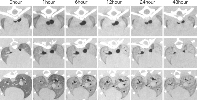
Methods: In this study, we examined temporal changes on postmortem chest CT images of a rat model. After acquiring antemortem images under isoflurane inhalation anesthesia, the rats were euthanized with a rapid intravenous injection of anesthetics. From immediately after death to 48 hours postmortem, chest images were acquired using small-animal CT. The 3D images were then evaluated on a workstation to measure the antemortem and postmortem air content in the lungs, trachea, and bronchi over time.
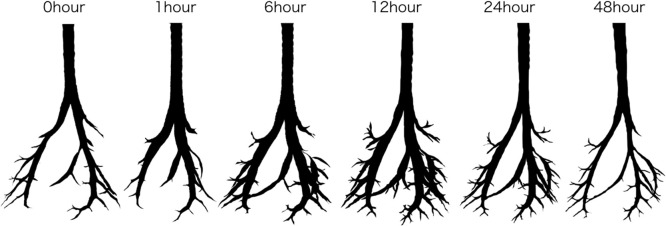
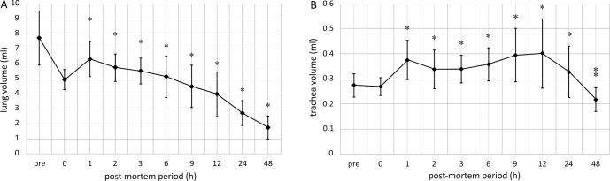
Results: The air content in the lungs decreased, but the air content of the trachea and bronchi temporarily increased 1-12 hours postmortem, then decreased at 48 hours postmortem. Therefore, the measurement of trachea and bronchi volumes on PMCT could be an objective way to estimate the time of death.
Conclusions: While the air content of the lungs decreased, the volume of the trachea and bronchi temporarily increased after death, indicating the potential to use such measurements to estimate time of death.


 分享
分享

 求助内容:
求助内容: 应助结果提醒方式:
 扫码关注我们
