{"title":"Idiopathic Unilateral Foveal Hemorrhage in a Young Woman Without Precipitating Factors: Evaluation with Optical Coherence Tomography Angiography.","authors":"Oguzhan Kilicarslan, Aslihan Yilmaz Cebi, Didar Ucar","doi":"10.14744/bej.2023.34976","DOIUrl":null,"url":null,"abstract":"<p><p>A 28-year-old young Caucasian female patient without a history of trauma or vascular disease presented with blurred vision and paracentral scotoma in her left eye. Fundus examination showed a small foveal hemorrhage in the superficial retinal layers. Initial visual acuity was 20/50 in the LE. After 2 weeks, visual acuity increased to 20/20, and hemorrhage was resolved in optical coherence tomography angiography (OCT-A) spontaneously. No vascular lesion was seen in any layer of the retina in OCT-A analysis.</p>","PeriodicalId":8740,"journal":{"name":"Beyoglu Eye Journal","volume":"8 2","pages":"134-138"},"PeriodicalIF":0.0000,"publicationDate":"2023-01-01","publicationTypes":"Journal Article","fieldsOfStudy":null,"isOpenAccess":false,"openAccessPdf":"https://ftp.ncbi.nlm.nih.gov/pub/pmc/oa_pdf/57/2b/BEJ-8-134.PMC10375203.pdf","citationCount":"0","resultStr":null,"platform":"Semanticscholar","paperid":null,"PeriodicalName":"Beyoglu Eye Journal","FirstCategoryId":"1085","ListUrlMain":"https://doi.org/10.14744/bej.2023.34976","RegionNum":0,"RegionCategory":null,"ArticlePicture":[],"TitleCN":null,"AbstractTextCN":null,"PMCID":null,"EPubDate":"","PubModel":"","JCR":"","JCRName":"","Score":null,"Total":0}
引用次数: 0
Abstract
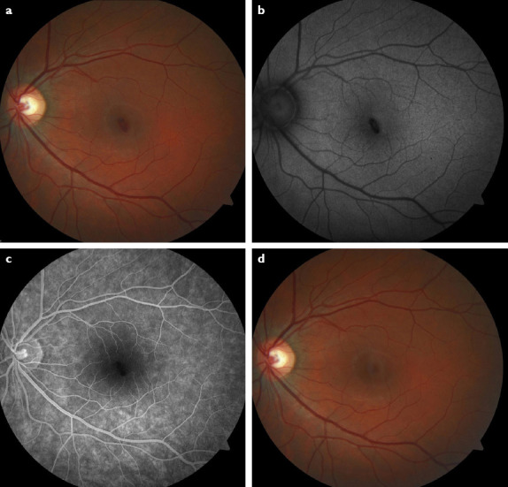
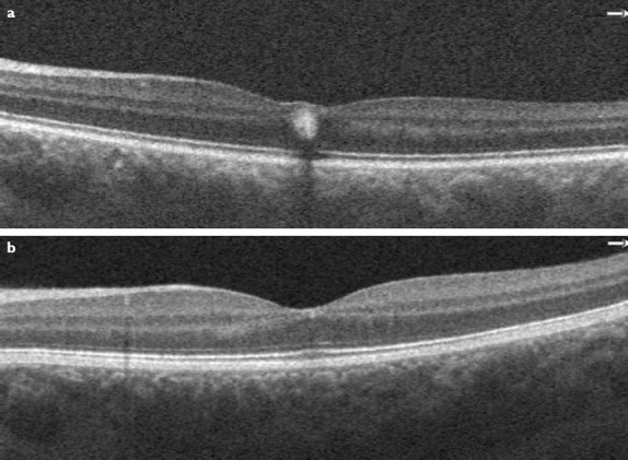
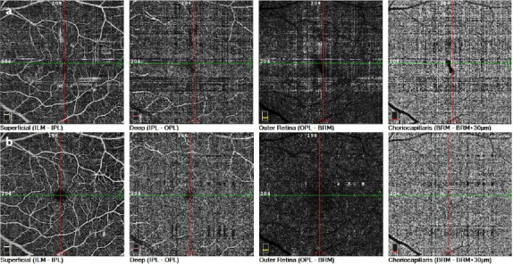
A 28-year-old young Caucasian female patient without a history of trauma or vascular disease presented with blurred vision and paracentral scotoma in her left eye. Fundus examination showed a small foveal hemorrhage in the superficial retinal layers. Initial visual acuity was 20/50 in the LE. After 2 weeks, visual acuity increased to 20/20, and hemorrhage was resolved in optical coherence tomography angiography (OCT-A) spontaneously. No vascular lesion was seen in any layer of the retina in OCT-A analysis.


 分享
分享

 求助内容:
求助内容: 应助结果提醒方式:
 扫码关注我们
