Stefan Georgiev, Manuel Ruiss, Andreea Dana-Fisus, Rainer A Leitgeb, Oliver Findl
{"title":"Comparison of corneal aberrations from anterior segment swept source OCT versus Placido-topography combined spectral domain OCT in cataract patients.","authors":"Stefan Georgiev, Manuel Ruiss, Andreea Dana-Fisus, Rainer A Leitgeb, Oliver Findl","doi":"10.1186/s40662-023-00348-z","DOIUrl":null,"url":null,"abstract":"<p><strong>Background: </strong>To comprehensively evaluate the agreement of component corneal aberrations from the newly updated wavefront analysis software of a swept-source optical coherence tomographer (SS-OCT) and a referential Placido-topography combined OCT device in elderly cataract patients.</p><p><strong>Methods: </strong>Retrospective study including 103 eyes from 103 elderly patients scheduled for cataract surgery that were measured on the same day with a SS-OCT (Heidelberg Engineering, Germany) device and a Placido-topography combined OCT device (CSO, Italy). Anterior, total, and posterior corneal wavefront aberrations were evaluated for their mean differences and limits of agreement (LoA) via Bland-Altman plots. Vector analysis was additionally employed to compare corneal astigmatism measurements in dioptric vector space.</p><p><strong>Results: </strong>Mean differences of all corneal aberrometric parameters did not exceed 0.05 μm. Total corneal aberrations were not significantly different from 0 except for vertical coma (- 0.04 μm; P = 0.003), spherical aberration (- 0.01 μm, P < 0.001), and root mean square (RMS) higher-order aberration (HOA) (0.03 μm, P = 0.04). The 95% LoA for total corneal aberration parameters between both devices were - 0.46 to 0.42 μm for horizontal astigmatism, - 0.37 to 0.41 μm for oblique astigmatism, - 0.19 to 0.17 μm for oblique trefoil, - 0.33 to 0.25 μm for vertical coma, - 0.20 to 0.22 μm for horizontal coma, - 0.22 to 0.20 μm for horizontal trefoil, - 0.11 to 0.08 μm for spherical aberration, and - 0.22 to 0.28 μm for RMS HOA. Vector analysis revealed no statistically significant mean differences for anterior, total, and posterior corneal astigmatism in dioptric vector space.</p><p><strong>Conclusion: </strong>In eyes undergoing cataract surgery with a regular elderly cornea, corneal wavefront analysis from the SS-OCT device showed functional equivalency to the reference device. Nevertheless, clinically relevant higher order aberration parameters should be interpreted with caution for surgical decision-making.</p>","PeriodicalId":73010,"journal":{"name":"","volume":"10 1","pages":"30"},"PeriodicalIF":0.0,"publicationDate":"2023-08-01","publicationTypes":"Journal Article","fieldsOfStudy":null,"isOpenAccess":false,"openAccessPdf":"https://www.ncbi.nlm.nih.gov/pmc/articles/PMC10392018/pdf/","citationCount":"0","resultStr":null,"platform":"Semanticscholar","paperid":null,"PeriodicalName":"","FirstCategoryId":"3","ListUrlMain":"https://doi.org/10.1186/s40662-023-00348-z","RegionNum":0,"RegionCategory":null,"ArticlePicture":[],"TitleCN":null,"AbstractTextCN":null,"PMCID":null,"EPubDate":"","PubModel":"","JCR":"","JCRName":"","Score":null,"Total":0}
引用次数: 0
Abstract
Background: To comprehensively evaluate the agreement of component corneal aberrations from the newly updated wavefront analysis software of a swept-source optical coherence tomographer (SS-OCT) and a referential Placido-topography combined OCT device in elderly cataract patients.
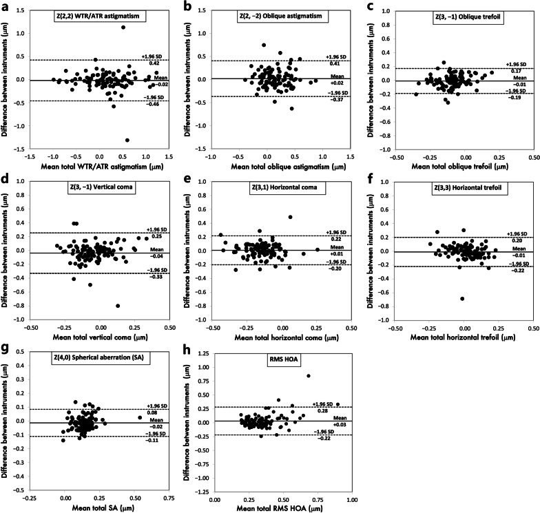
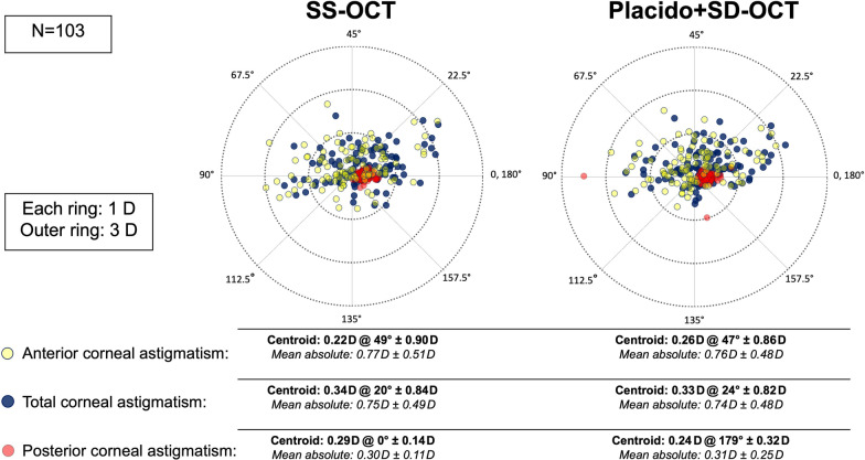
Methods: Retrospective study including 103 eyes from 103 elderly patients scheduled for cataract surgery that were measured on the same day with a SS-OCT (Heidelberg Engineering, Germany) device and a Placido-topography combined OCT device (CSO, Italy). Anterior, total, and posterior corneal wavefront aberrations were evaluated for their mean differences and limits of agreement (LoA) via Bland-Altman plots. Vector analysis was additionally employed to compare corneal astigmatism measurements in dioptric vector space.
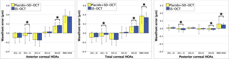
Results: Mean differences of all corneal aberrometric parameters did not exceed 0.05 μm. Total corneal aberrations were not significantly different from 0 except for vertical coma (- 0.04 μm; P = 0.003), spherical aberration (- 0.01 μm, P < 0.001), and root mean square (RMS) higher-order aberration (HOA) (0.03 μm, P = 0.04). The 95% LoA for total corneal aberration parameters between both devices were - 0.46 to 0.42 μm for horizontal astigmatism, - 0.37 to 0.41 μm for oblique astigmatism, - 0.19 to 0.17 μm for oblique trefoil, - 0.33 to 0.25 μm for vertical coma, - 0.20 to 0.22 μm for horizontal coma, - 0.22 to 0.20 μm for horizontal trefoil, - 0.11 to 0.08 μm for spherical aberration, and - 0.22 to 0.28 μm for RMS HOA. Vector analysis revealed no statistically significant mean differences for anterior, total, and posterior corneal astigmatism in dioptric vector space.
Conclusion: In eyes undergoing cataract surgery with a regular elderly cornea, corneal wavefront analysis from the SS-OCT device showed functional equivalency to the reference device. Nevertheless, clinically relevant higher order aberration parameters should be interpreted with caution for surgical decision-making.


 分享
分享
 求助内容:
求助内容: 应助结果提醒方式:
 扫码关注我们
