Hemn H Kaka Ali, Dana T Gharib, Marwan N Hassan, Ari M Abdullah, Deari A Ismaeil, Omar H Ghalib Hawramy, Dlshad H Ahmed, Dilan S Hiwa, Berun A Abdalla, Fahmi H Kakamad
{"title":"Biliary tree traumatic neuroma following laparoscopic cholecystectomy: A case report and literature review.","authors":"Hemn H Kaka Ali, Dana T Gharib, Marwan N Hassan, Ari M Abdullah, Deari A Ismaeil, Omar H Ghalib Hawramy, Dlshad H Ahmed, Dilan S Hiwa, Berun A Abdalla, Fahmi H Kakamad","doi":"10.3892/mi.2023.97","DOIUrl":null,"url":null,"abstract":"<p><p>Laparoscopic cholecystectomy has been found to be associated with the development of traumatic neuromas on rare occasions. The present study reports a rare case of post-cholecystectomy biliary tree traumatic neuroma. Herein, a 47-year-old female with a history of laparoscopic cholecystectomy presented with upper abdominal pain and anorexia. Upon an examination, a yellow discoloration of the sclera was observed. Magnetic resonance cholangiopancreatography revealed a dilated proximal bile duct and mild dilatation of the intrahepatic biliary tree due to a stricture. Intraoperatively, a hard bile duct mass was observed with multiple enlarged lymph nodes in the peri-hepatic region. The patient was initially suspected to have bile duct cancer; however, a histopathological analysis of the resected mass revealed a bile duct traumatic neuroma. Biliary traumatic neuromas may be underestimated since they often remain asymptomatic. It is unfortunate that, as traumatic neuromas often lack distinguishing characteristics, no particular radiological findings for traumatic neuromas of the bile duct have been described to date, at least to the best of our knowledge. The rarity of this condition, combined with the absence of a standardized diagnostic modality, renders its diagnosis difficult and can even lead to misdiagnosis as biliary cancer.</p>","PeriodicalId":74161,"journal":{"name":"Medicine international","volume":"3 4","pages":"37"},"PeriodicalIF":0.0000,"publicationDate":"2023-07-01","publicationTypes":"Journal Article","fieldsOfStudy":null,"isOpenAccess":false,"openAccessPdf":"https://www.ncbi.nlm.nih.gov/pmc/articles/PMC10391593/pdf/","citationCount":"0","resultStr":null,"platform":"Semanticscholar","paperid":null,"PeriodicalName":"Medicine international","FirstCategoryId":"1085","ListUrlMain":"https://doi.org/10.3892/mi.2023.97","RegionNum":0,"RegionCategory":null,"ArticlePicture":[],"TitleCN":null,"AbstractTextCN":null,"PMCID":null,"EPubDate":"","PubModel":"","JCR":"","JCRName":"","Score":null,"Total":0}
引用次数: 0
Abstract
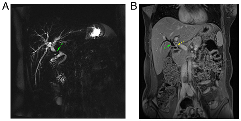
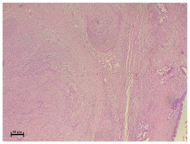
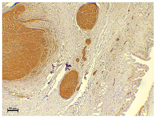
Laparoscopic cholecystectomy has been found to be associated with the development of traumatic neuromas on rare occasions. The present study reports a rare case of post-cholecystectomy biliary tree traumatic neuroma. Herein, a 47-year-old female with a history of laparoscopic cholecystectomy presented with upper abdominal pain and anorexia. Upon an examination, a yellow discoloration of the sclera was observed. Magnetic resonance cholangiopancreatography revealed a dilated proximal bile duct and mild dilatation of the intrahepatic biliary tree due to a stricture. Intraoperatively, a hard bile duct mass was observed with multiple enlarged lymph nodes in the peri-hepatic region. The patient was initially suspected to have bile duct cancer; however, a histopathological analysis of the resected mass revealed a bile duct traumatic neuroma. Biliary traumatic neuromas may be underestimated since they often remain asymptomatic. It is unfortunate that, as traumatic neuromas often lack distinguishing characteristics, no particular radiological findings for traumatic neuromas of the bile duct have been described to date, at least to the best of our knowledge. The rarity of this condition, combined with the absence of a standardized diagnostic modality, renders its diagnosis difficult and can even lead to misdiagnosis as biliary cancer.


 分享
分享

 求助内容:
求助内容: 应助结果提醒方式:
 扫码关注我们
